 Movie
Movie Controller

[English] 日本語
 Yorodumi
- PDB-1szj: STRUCTURE OF HOLO-GLYCERALDEHYDE-3-PHOSPHATE-DEHYDROGENASE FROM P... -
+ Open data

- Basic information
| Entry | Database: PDB / ID: 1szj | ||||||
|---|---|---|---|---|---|---|---|
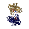
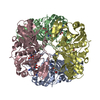
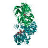
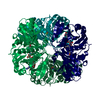
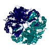
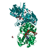
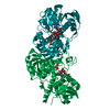
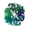
| Title | STRUCTURE OF HOLO-GLYCERALDEHYDE-3-PHOSPHATE-DEHYDROGENASE FROM PALINURUS VERSICOLOR REFINED 2.0 ANGSTROM RESOLUTION | ||||||
 Components Components | D-GLYCERALDEHYDE-3-PHOSPHATE-DEHYDROGENASE | ||||||
 Keywords Keywords | OXIDOREDUCTASE / D-GLYCERALDEHYDE-3-PHOSPHATE-DEHYDROGENASE / MOLECULAR SYMMETRY / ALLOSTERISM | ||||||
| Function / homology |  Function and homology information Function and homology informationglyceraldehyde-3-phosphate metabolic process / glyceraldehyde-3-phosphate dehydrogenase (phosphorylating) / glyceraldehyde-3-phosphate dehydrogenase (NAD+) (phosphorylating) activity / glycolytic process / glucose metabolic process / NAD binding / NADP binding / cytosol Similarity search - Function | ||||||
| Biological species |  Palinurus versicolor (painted spiny lobster) Palinurus versicolor (painted spiny lobster) | ||||||
| Method |  X-RAY DIFFRACTION / SYNCHROTRON / MOLECULAR REPLACEMENT / Resolution: 2 Å X-RAY DIFFRACTION / SYNCHROTRON / MOLECULAR REPLACEMENT / Resolution: 2 Å | ||||||
 Authors Authors | Song, S. / Li, J. / Lin, Z. | ||||||
 Citation Citation | Journal: J.Mol.Biol. / Year: 1983 Title: Preliminary crystallographic studies of lobster D-glyceraldehyde-3-phosphate dehydrogenase and the modified enzyme carrying the fluorescent derivative. Authors: Song, S.Y. / Gao, Y.G. / Zhou, J.M. / Tsou, C.L. #1: Journal: Arch.Biochem.Biophys. / Year: 1993Title: Structure of D-Glyceraldehyde-3-Phosphate Dehydrogenase from Palinurus Versicolor Carrying the Fluorescent Nad Derivatives at 2.7 A Resolution Authors: Lin, Z.J. / Li, J. / Zhang, F.M. / Song, S.Y. / Yang, J. / Liang, S.J. / Tsou, C.L. | ||||||
| History |
|
- Structure visualization
| Structure viewer | Molecule: MolmilJmol/JSmol |
|---|
- Downloads & links
-Download
| PDBx/mmCIF format | 1szj.cif.gz | 146.9 KB | Display | PDBx/mmCIF format |
|---|---|---|---|---|
| PDB format | pdb1szj.ent.gz | 114.6 KB | Display | PDB format |
| PDBx/mmJSON format | 1szj.json.gz | Tree view | PDBx/mmJSON format | |
| Others |  Other downloads Other downloads |
-Validation report
| Arichive directory | https://data.pdbj.org/pub/pdb/validation_reports/sz/1szjftp://data.pdbj.org/pub/pdb/validation_reports/sz/1szj | HTTPS FTP |
|---|
-Related structure data
| Similar structure data |
|---|


-Links
 PDBj
PDBj
- Assembly
| Deposited unit | 
| ||||||||
|---|---|---|---|---|---|---|---|---|---|
| 1 | 
| ||||||||
| Unit cell |
| ||||||||
| Noncrystallographic symmetry (NCS) | NCS oper: (Code: given Matrix: (-0.99997, -0.00521, -0.0049), Vector: |
-Components
| #1: Protein | Mass: 35767.004 Da / Num. of mol.: 2 / Fragment: NAD+ BINDING DOMAIN AND CATALYTIC DOMAIN / Source method: isolated from a natural source / Details: COMPLEX WITH NAD+ Source: (natural) Palinurus versicolor (painted spiny lobster)Organ: TAIL / Tissue: TAIL MUSCLE References: UniProt: P56649, glyceraldehyde-3-phosphate dehydrogenase (phosphorylating) #2: Chemical | ChemComp-SO4 /   Mass: 96.063 Da / Num. of mol.: 4 / Source method: obtained synthetically / Formula: SO4 Mass: 96.063 Da / Num. of mol.: 4 / Source method: obtained synthetically / Formula: SO4#3: Chemical |   Mass: 663.425 Da / Num. of mol.: 2 / Source method: obtained synthetically / Formula: C21H27N7O14P2 / Comment: NAD*YM Mass: 663.425 Da / Num. of mol.: 2 / Source method: obtained synthetically / Formula: C21H27N7O14P2 / Comment: NAD*YM#4: Water | ChemComp-HOH / |  Mass: 18.015 Da / Num. of mol.: 366 / Source method: isolated from a natural source / Formula: H2O Mass: 18.015 Da / Num. of mol.: 366 / Source method: isolated from a natural source / Formula: H2O |
|---|
-Experimental details
-Experiment
| Experiment | Method: X-RAY DIFFRACTION / Number of used crystals: 1 |
|---|
- Sample preparation
| Crystal | Density Matthews: 3.27 Å3/Da / Density % sol: 62 % Description: THE 2.7 ANGSTROM STRUCTURE OF IRR-GAPDH FROM PALINURUS VERSICOLOR WAS DETERMINED BY MOLECULAR REPLACEMENT USING HOMARUS AMERICANUS HOLO-GAPDH AS A SEARCH MODEL. |
|---|---|
| Crystal grow | Temperature: 280 K / pH: 6.1 Details: THE PROTEIN SOLUTIONS CONTAINED 0.5MM NAD+, 1.0MM EDTA, 1.6M AMMONIUM SULPHATE IN 0.1M PHOSPHATE BUFFER (PH 6.1) AND AN ENZYME CONCENTRATION OF 8MG/ML; THE SOLUTION IN RESERVOIR CONTAINED 2. ...Details: THE PROTEIN SOLUTIONS CONTAINED 0.5MM NAD+, 1.0MM EDTA, 1.6M AMMONIUM SULPHATE IN 0.1M PHOSPHATE BUFFER (PH 6.1) AND AN ENZYME CONCENTRATION OF 8MG/ML; THE SOLUTION IN RESERVOIR CONTAINED 2.7M AMMONIUM SULPHATE IN SAME BUFFER. THE CRYSTALLIZATION WAS CARRIED OUT AT 280K. |
-Data collection
| Diffraction | Mean temperature: 280 K |
|---|---|
| Diffraction source | Source: SYNCHROTRON / Site: Photon Factory  / Beamline: BL-6A / Wavelength: 1 / Beamline: BL-6A / Wavelength: 1 |
| Detector | Type: WEISSENBERG / Detector: DIFFRACTOMETER / Date: May 1, 1994 |
| Radiation | Monochromator: SI(111) / Monochromatic (M) / Laue (L): M / Scattering type: x-ray |
| Radiation wavelength | Wavelength: 1 Å / Relative weight: 1 |
| Reflection | Resolution: 1.7→100 Å / Num. obs: 84900 / % possible obs: 83.5 % / Observed criterion σ(I): 0 / Redundancy: 1.4 % / Biso Wilson estimate: 21.93 Å2 / Rmerge(I) obs: 0.069 / Net I/σ(I): 3.9 |
| Reflection shell | Resolution: 1.7→1.74 Å / Redundancy: 1.1 % / Rmerge(I) obs: 0.069 / Mean I/σ(I) obs: 1.6 / % possible all: 56.5 |
- Processing
| Software |
| ||||||||||||||||||||||||||||||||||||||||||||||||||||||||||||
|---|---|---|---|---|---|---|---|---|---|---|---|---|---|---|---|---|---|---|---|---|---|---|---|---|---|---|---|---|---|---|---|---|---|---|---|---|---|---|---|---|---|---|---|---|---|---|---|---|---|---|---|---|---|---|---|---|---|---|---|---|---|
| Refinement | Method to determine structure: MOLECULAR REPLACEMENT Starting model: HOMARUS AMERICANUS HOLO-GAPDH Resolution: 2→6 Å / Data cutoff low absF: 50 / σ(F): 3 Details: DURING THE REFINEMENT, NO NCS RESTRAINTS WERE USED.
| ||||||||||||||||||||||||||||||||||||||||||||||||||||||||||||
| Displacement parameters | Biso mean: 32.39 Å2 | ||||||||||||||||||||||||||||||||||||||||||||||||||||||||||||
| Refine analyze | Luzzati coordinate error obs: 0.22 Å / Luzzati d res low obs: 6 Å | ||||||||||||||||||||||||||||||||||||||||||||||||||||||||||||
| Refinement step | Cycle: LAST / Resolution: 2→6 Å
| ||||||||||||||||||||||||||||||||||||||||||||||||||||||||||||
| Refine LS restraints |
| ||||||||||||||||||||||||||||||||||||||||||||||||||||||||||||
| LS refinement shell | Resolution: 1.98→2.09 Å
| ||||||||||||||||||||||||||||||||||||||||||||||||||||||||||||
| Xplor file |
|
