 Movie
Movie Controller

[English] 日本語
 Yorodumi
- PDB-2mcp: REFINED CRYSTAL STRUCTURE OF THE MC/PC603 FAB-PHOSPHOCHOLINE COMP... -
+ Open data

- Basic information
| Entry | Database: PDB / ID: 2mcp | |||||||||
|---|---|---|---|---|---|---|---|---|---|---|
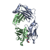
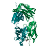
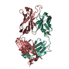
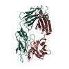
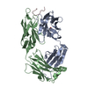
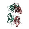
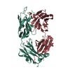
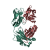
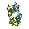
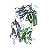
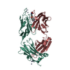
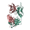
| Title | REFINED CRYSTAL STRUCTURE OF THE MC/PC603 FAB-PHOSPHOCHOLINE COMPLEX AT 3.1 ANGSTROMS RESOLUTION | |||||||||
 Components Components |
| |||||||||
 Keywords Keywords | IMMUNE SYSTEM / IMMUNOGLOBULIN | |||||||||
| Function / homology |  Function and homology information Function and homology informationimmunoglobulin mediated immune response / immunoglobulin complex / antigen binding / extracellular region Similarity search - Function | |||||||||
| Biological species |  | |||||||||
| Method |  X-RAY DIFFRACTION / Resolution: 3.1 Å X-RAY DIFFRACTION / Resolution: 3.1 Å | |||||||||
 Authors Authors | Padlan, E.A. / Cohen, G.H. / Davies, D.R. | |||||||||
 Citation Citation | Journal: To be Published Title: Refined Crystal Structure of the Mc/Pc603 Fab-Phosphocholine Complex at 3.1 Angstroms Resolution Authors: Padlan, E.A. / Cohen, G.H. / Davies, D.R. #1: Journal: Ann.Immunol.(Paris),Sect.C / Year: 1985Title: On the Specificity of Antibody(Slash)Antigen Interactions. Phosphocholine Binding to Mc/Pc603 and the Correlation of Three-Dimensional Structure and Sequence Data Authors: Padlan, E.A. / Cohen, G.H. / Davies, D.R. #2: Journal: The Immune System. Genes,Receptors,Signals. Proceedings of the 1974 I.C.N.-U.C.L.A. Symposium on Molecular BiologyYear: 1974 Title: The Three-Dimensional Structure of the Antigen Binding Site of Mc/Pc 603 Protein Authors: Padlan, E.A. / Segal, D.M. / Cohen, G.H. / Davies, D.R. / Rudikoff, S. / Potter, M. #3: Journal: Proc.Natl.Acad.Sci.USA / Year: 1974Title: The Three-Dimensional Structure of a Phosphorylcholine-Binding Mouse Immunoglobulin Fab and the Nature of the Antigen Binding Site Authors: Segal, D.M. / Padlan, E.A. / Cohen, G.H. / Rudikoff, S. / Potter, M. / Davies, D.R. #4: Journal: Nature New Biol. / Year: 1973Title: Structure at 4.5 Angstroms Resolution of a Phosphorylcholine-Binding Fab Structure at 4.5 Angstroms Resolution of a Phosphorylcholine-Binding Fab Authors: Padlan, E.A. / Segal, D.M. / Spande, T.F. / Rudikoff, D.R.Davies S. / Potter, M. | |||||||||
| History |
|
- Structure visualization
| Structure viewer | Molecule: MolmilJmol/JSmol |
|---|
- Downloads & links
-Download
| PDBx/mmCIF format | 2mcp.cif.gz | 94.5 KB | Display | PDBx/mmCIF format |
|---|---|---|---|---|
| PDB format | pdb2mcp.ent.gz | 72.5 KB | Display | PDB format |
| PDBx/mmJSON format | 2mcp.json.gz | Tree view | PDBx/mmJSON format | |
| Others |  Other downloads Other downloads |
-Validation report
| Arichive directory | https://data.pdbj.org/pub/pdb/validation_reports/mc/2mcpftp://data.pdbj.org/pub/pdb/validation_reports/mc/2mcp | HTTPS FTP |
|---|
-Related structure data
| Similar structure data |
|---|


-Links
 PDBj
PDBj

- Assembly
| Deposited unit | 
| ||||||||
|---|---|---|---|---|---|---|---|---|---|
| 1 |
| ||||||||
| Unit cell |
| ||||||||
| Atom site foot note | 1: RESIDUES PRO L 8, PRO L 101, PRO L 147, PRO H 143 AND PRO H 155 ARE CIS PROLINES. |
-Components
| #1: Antibody | Mass: 24113.584 Da / Num. of mol.: 1 Source method: isolated from a genetically manipulated source Source: (gene. exp.) |
|---|---|
| #2: Antibody | Mass: 24319.266 Da / Num. of mol.: 1 Source method: isolated from a genetically manipulated source Source: (gene. exp.) |
| #3: Chemical | ChemComp-PC /   Mass: 184.151 Da / Num. of mol.: 1 / Source method: obtained synthetically / Formula: C5H15NO4P Mass: 184.151 Da / Num. of mol.: 1 / Source method: obtained synthetically / Formula: C5H15NO4P |
| Has protein modification | Y |
-Experimental details
-Experiment
| Experiment | Method: X-RAY DIFFRACTION |
|---|
- Sample preparation
| Crystal | Density Matthews: 4.78 Å3/Da / Density % sol: 74.25 % |
|---|
-Data collection
| Radiation | Scattering type: x-ray |
|---|---|
| Radiation wavelength | Relative weight: 1 |
- Processing
| Refinement | Rfactor Rwork: 0.185 / Highest resolution: 3.1 Å | ||||||||||||
|---|---|---|---|---|---|---|---|---|---|---|---|---|---|
| Refinement step | Cycle: LAST / Highest resolution: 3.1 Å
|
