 Movie
Movie Controller

[English] 日本語
 Yorodumi
- PDB-1a8j: IMMUNOGLOBULIN LAMBDA LIGHT CHAIN DIMER (MCG) COMPLEX WITH ASPARTAME -
+ Open data

- Basic information
| Entry | Database: PDB / ID: 1a8j | |||||||||
|---|---|---|---|---|---|---|---|---|---|---|
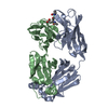
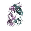
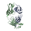
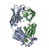
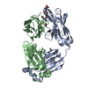
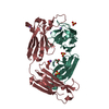
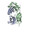
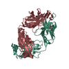
| Title | IMMUNOGLOBULIN LAMBDA LIGHT CHAIN DIMER (MCG) COMPLEX WITH ASPARTAME | |||||||||
 Components Components | IMMUNOGLOBULIN LAMBDA LIGHT CHAIN DIMER (MCG) | |||||||||
 Keywords Keywords | IMMUNOGLOBULIN / ASPARTAME / LIGAND BINDING / IMMUNOTHERAPEUTIC AGENT / SURROGATE RECEPTOR / MCG(HUMAN) BENCE-JONES DIMER | |||||||||
| Function / homology |  Function and homology information Function and homology informationCD22 mediated BCR regulation / Fc epsilon receptor (FCERI) signaling / Classical antibody-mediated complement activation / Initial triggering of complement / FCGR activation / Role of LAT2/NTAL/LAB on calcium mobilization / immunoglobulin complex / Role of phospholipids in phagocytosis / Scavenging of heme from plasma / antigen binding ...CD22 mediated BCR regulation / Fc epsilon receptor (FCERI) signaling / Classical antibody-mediated complement activation / Initial triggering of complement / FCGR activation / Role of LAT2/NTAL/LAB on calcium mobilization / immunoglobulin complex / Role of phospholipids in phagocytosis / Scavenging of heme from plasma / antigen binding / FCERI mediated Ca+2 mobilization / FCGR3A-mediated IL10 synthesis / Regulation of Complement cascade / Antigen activates B Cell Receptor (BCR) leading to generation of second messengers / Cell surface interactions at the vascular wall / FCGR3A-mediated phagocytosis / FCERI mediated MAPK activation / Regulation of actin dynamics for phagocytic cup formation / Immunoregulatory interactions between a Lymphoid and a non-Lymphoid cell / FCERI mediated NF-kB activation / Potential therapeutics for SARS / adaptive immune response / immune response / extracellular region / plasma membrane Similarity search - Function | |||||||||
| Biological species |  Homo sapiens (human) Homo sapiens (human) | |||||||||
| Method |  X-RAY DIFFRACTION / DIFFERENCE MAP / Resolution: 2.7 Å X-RAY DIFFRACTION / DIFFERENCE MAP / Resolution: 2.7 Å | |||||||||
 Authors Authors | Edmundson, A.B. / Manion, C.V. | |||||||||
 Citation Citation | Journal: Clin.Pharmacol.Ther. / Year: 1998 Title: Treatment of osteoarthritis with aspartame. Authors: Edmundson, A.B. / Manion, C.V. #1: Journal: J.Mol.Biol. / Year: 1989Title: Three-Dimensional Structure of a Light Chain Dimer Crystallized in Water. Conformational Flexibility of a Molecule in Two Crystal Forms Authors: Ely, K.R. / Herron, J.N. / Harker, M. / Edmundson, A.B. | |||||||||
| History |
|
- Structure visualization
| Structure viewer | Molecule: MolmilJmol/JSmol |
|---|
- Downloads & links
-Download
| PDBx/mmCIF format | 1a8j.cif.gz | 83.1 KB | Display | PDBx/mmCIF format |
|---|---|---|---|---|
| PDB format | pdb1a8j.ent.gz | 61.8 KB | Display | PDB format |
| PDBx/mmJSON format | 1a8j.json.gz | Tree view | PDBx/mmJSON format | |
| Others |  Other downloads Other downloads |
-Validation report
| Arichive directory | https://data.pdbj.org/pub/pdb/validation_reports/a8/1a8jftp://data.pdbj.org/pub/pdb/validation_reports/a8/1a8j | HTTPS FTP |
|---|
-Related structure data
| Similar structure data |
|---|


-Links
 PDBj
PDBj


- Assembly
| Deposited unit | 
| ||||||||
|---|---|---|---|---|---|---|---|---|---|
| 1 |
| ||||||||
| Unit cell |
|
-Components
| #1: Antibody | Mass: 22833.064 Da / Num. of mol.: 2 / Source method: isolated from a natural source Details: FOUND IN THE URINE OF PATIENT MCG WITH A MULTIPLE MYELOMA AND AMYLOIDOSIS Source: (natural) Homo sapiens (human) / References: UniProt: P01709#2: Chemical |   Mass: 294.303 Da / Num. of mol.: 2 / Source method: obtained synthetically / Formula: C14H18N2O5 Mass: 294.303 Da / Num. of mol.: 2 / Source method: obtained synthetically / Formula: C14H18N2O5Has protein modification | Y | |
|---|
-Experimental details
-Experiment
| Experiment | Method: X-RAY DIFFRACTION / Number of used crystals: 1 |
|---|
- Sample preparation
| Crystal | Density Matthews: 3.07 Å3/Da / Density % sol: 61 % | ||||||||||||||||||||
|---|---|---|---|---|---|---|---|---|---|---|---|---|---|---|---|---|---|---|---|---|---|
| Crystal grow | pH: 6.2 Details: PROTEIN WAS CRYSTALLIZED IN FRESH 1.9M AMMONIUM SULFATE BUFFERED AT PH 6.2 WITH PHOSPHATE AND THE MOTHER LIQUOR WAS SATURATED WITH ASPARTAME. | ||||||||||||||||||||
| Crystal grow | *PLUS Method: unknown | ||||||||||||||||||||
| Components of the solutions | *PLUS
|
-Data collection
| Diffraction | Mean temperature: 286 K |
|---|---|
| Diffraction source | Source: SEALED TUBE / Type: OTHER / Wavelength: 1.5418 |
| Detector | Type: SIEMENS / Detector: DIFFRACTOMETER / Date: Nov 1, 1988 |
| Radiation | Monochromator: NI FILTER / Monochromatic (M) / Laue (L): M / Scattering type: x-ray |
| Radiation wavelength | Wavelength: 1.5418 Å / Relative weight: 1 |
| Reflection | Highest resolution: 2.7 Å / Num. obs: 10570 / % possible obs: 62 % / Observed criterion σ(I): 2 / Biso Wilson estimate: 17 Å2 / Net I/σ(I): 20 |
| Reflection shell | Resolution: 2.7→2.82 Å / Mean I/σ(I) obs: 7 / % possible all: 30 |
- Processing
| Software |
| ||||||||||||||||||||||||||||||||||||||||||||||||||||||||||||
|---|---|---|---|---|---|---|---|---|---|---|---|---|---|---|---|---|---|---|---|---|---|---|---|---|---|---|---|---|---|---|---|---|---|---|---|---|---|---|---|---|---|---|---|---|---|---|---|---|---|---|---|---|---|---|---|---|---|---|---|---|---|
| Refinement | Method to determine structure: DIFFERENCE MAP / Resolution: 2.7→8 Å / Data cutoff high absF: 1000000 / Data cutoff low absF: 0.001 / Cross valid method: THROUGHOUT / σ(F): 0
| ||||||||||||||||||||||||||||||||||||||||||||||||||||||||||||
| Displacement parameters | Biso mean: 17 Å2 | ||||||||||||||||||||||||||||||||||||||||||||||||||||||||||||
| Refinement step | Cycle: LAST / Resolution: 2.7→8 Å
| ||||||||||||||||||||||||||||||||||||||||||||||||||||||||||||
| Refine LS restraints |
| ||||||||||||||||||||||||||||||||||||||||||||||||||||||||||||
| LS refinement shell | Resolution: 2.7→2.82 Å / Total num. of bins used: 8
| ||||||||||||||||||||||||||||||||||||||||||||||||||||||||||||
| Xplor file |
| ||||||||||||||||||||||||||||||||||||||||||||||||||||||||||||
| Software | *PLUS Name: X-PLOR / Version: 3.8 / Classification: refinement | ||||||||||||||||||||||||||||||||||||||||||||||||||||||||||||
| Refine LS restraints | *PLUS
|
