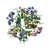
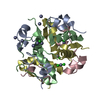
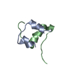
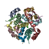
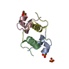
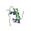
SHEET THE SHEET STRUCTURE OF THIS MOLECULE IS BIFURCATED. IN ORDER TO REPRESENT THIS FEATURE IN ... SHEET THE SHEET STRUCTURE OF THIS MOLECULE IS BIFURCATED. IN ORDER TO REPRESENT THIS FEATURE IN THE SHEET RECORDS BELOW, TWO SHEETS ARE DEFINED.
Mass: 18.015 Da / Num. of mol.: 360 / Source method: isolated from a natural source / Formula: H2O
-
Details
Compound details
ENGINEERED RESIDUE IN CHAIN A, CYS 110 TO LEU ENGINEERED RESIDUE IN CHAIN A, GLU 111 TO GLN ...ENGINEERED RESIDUE IN CHAIN A, CYS 110 TO LEU ENGINEERED RESIDUE IN CHAIN A, GLU 111 TO GLN ENGINEERED RESIDUE IN CHAIN A, CYS 171 TO SER ENGINEERED RESIDUE IN CHAIN A, CYS 178 TO ALA ENGINEERED RESIDUE IN CHAIN A, CYS 257 TO VAL ENGINEERED RESIDUE IN CHAIN A, CYS 414 TO LEU ENGINEERED RESIDUE IN CHAIN A, CYS 573 TO ASN ENGINEERED RESIDUE IN CHAIN A, CYS 590 TO SER ENGINEERED RESIDUE IN CHAIN A, CYS 789 TO SER ENGINEERED RESIDUE IN CHAIN A, CYS 812 TO ALA ENGINEERED RESIDUE IN CHAIN A, CYS 819 TO ALA ENGINEERED RESIDUE IN CHAIN A, CYS 904 TO SER ENGINEERED RESIDUE IN CHAIN A, CYS 966 TO ASN ENGINEERED RESIDUE IN CHAIN A, CYS 974 TO ALA ENGINEERED RESIDUE IN CHAIN B, CYS 110 TO LEU ENGINEERED RESIDUE IN CHAIN B, GLU 111 TO GLN ENGINEERED RESIDUE IN CHAIN B, CYS 171 TO SER ENGINEERED RESIDUE IN CHAIN B, CYS 178 TO ALA ENGINEERED RESIDUE IN CHAIN B, CYS 257 TO VAL ENGINEERED RESIDUE IN CHAIN B, CYS 414 TO LEU ENGINEERED RESIDUE IN CHAIN B, CYS 573 TO ASN ENGINEERED RESIDUE IN CHAIN B, CYS 590 TO SER ENGINEERED RESIDUE IN CHAIN B, CYS 789 TO SER ENGINEERED RESIDUE IN CHAIN B, CYS 812 TO ALA ENGINEERED RESIDUE IN CHAIN B, CYS 819 TO ALA ENGINEERED RESIDUE IN CHAIN B, CYS 904 TO SER ENGINEERED RESIDUE IN CHAIN B, CYS 966 TO ASN ENGINEERED RESIDUE IN CHAIN B, CYS 974 TO ALA
Has protein modification
Y
-
Experimental details
-
Experiment
Experiment
Method: X-RAY DIFFRACTION / Number of used crystals: 1
-
Sample preparation
Crystal
Density Matthews: 3.87 Å3/Da / Density % sol: 67.93 % / Description: NONE
Resolution: 2.8→29.85 Å / Cor.coef. Fo:Fc: 0.949 / Cor.coef. Fo:Fc free: 0.918 / SU B: 9.117 / SU ML: 0.182 / Cross valid method: THROUGHOUT / ESU R: 0.471 / ESU R Free: 0.279 / Stereochemistry target values: MAXIMUM LIKELIHOOD / Details: HYDROGENS HAVE BEEN ADDED IN THE RIDING POSITIONS
Rfactor
Num. reflection
% reflection
Selection details
Rfree
0.22
4422
5 %
RANDOM
Rwork
0.17
-
-
-
obs
0.172
83937
99.7 %
-
Solvent computation
Ion probe radii: 0.8 Å / Shrinkage radii: 0.8 Å / VDW probe radii: 1.2 Å / Solvent model: MASK
 Movie
Movie Controller

 Yorodumi
 Open data
 Basic information
 Components
 Keywords
 Function and homology information
 HOMO SAPIENS (human)
 X-RAY DIFFRACTION /
  Authors
 Citation
 Structure visualization
 Downloads & links
 Other downloads


 PDBj
PDBj


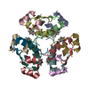
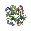
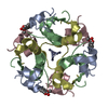
 Assembly


 Mass: 65.409 Da / Num. of mol.: 2 / Source method: obtained synthetically / Formula: Zn
 Mass: 88.105 Da / Num. of mol.: 8 / Source method: obtained synthetically / Formula: C4H8O2
 Sample preparation
 / Beamline: 19-ID / Wavelength: 1.548
  Processing