 Movie
Movie Controller

[English] 日本語
 Yorodumi
- PDB-1xw7: Diabetes-Associated Mutations in Human Insulin: Crystal Structure... -
+ Open data

- Basic information
| Entry | Database: PDB / ID: 1xw7 | ||||||
|---|---|---|---|---|---|---|---|
| Title | Diabetes-Associated Mutations in Human Insulin: Crystal Structure and Photo-Cross-Linking Studies of A-Chain Variant Insulin Wakayama | ||||||
 Components Components | (Insulin) x 2 | ||||||
 Keywords Keywords | HORMONE/GROWTH FACTOR / Leu-A30insulin / protein unfolding / insulin receptor / HORMONE-GROWTH FACTOR COMPLEX | ||||||
| Function / homology |  Function and homology information Function and homology informationnegative regulation of glycogen catabolic process / : / negative regulation of fatty acid metabolic process / Signaling by Insulin receptor / negative regulation of feeding behavior / IRS activation / Insulin processing / regulation of protein secretion / positive regulation of peptide hormone secretion / negative regulation of acute inflammatory response ...negative regulation of glycogen catabolic process / : / negative regulation of fatty acid metabolic process / Signaling by Insulin receptor / negative regulation of feeding behavior / IRS activation / Insulin processing / regulation of protein secretion / positive regulation of peptide hormone secretion / negative regulation of acute inflammatory response / Regulation of gene expression in beta cells / positive regulation of respiratory burst / alpha-beta T cell activation / Synthesis, secretion, and deacylation of Ghrelin / negative regulation of protein secretion / positive regulation of dendritic spine maintenance / negative regulation of gluconeogenesis / fatty acid homeostasis / positive regulation of glycogen biosynthetic process / positive regulation of insulin receptor signaling pathway / Signal attenuation / FOXO-mediated transcription of oxidative stress, metabolic and neuronal genes / negative regulation of lipid catabolic process / negative regulation of respiratory burst involved in inflammatory response / positive regulation of lipid biosynthetic process / negative regulation of oxidative stress-induced intrinsic apoptotic signaling pathway / nitric oxide-cGMP-mediated signaling / regulation of protein localization to plasma membrane / positive regulation of nitric-oxide synthase activity / transport vesicle / Insulin receptor recycling / COPI-mediated anterograde transport / positive regulation of brown fat cell differentiation / negative regulation of reactive oxygen species biosynthetic process / insulin-like growth factor receptor binding / NPAS4 regulates expression of target genes / neuron projection maintenance / positive regulation of mitotic nuclear division / endoplasmic reticulum-Golgi intermediate compartment membrane / Insulin receptor signalling cascade / positive regulation of glycolytic process / endosome lumen / positive regulation of cytokine production / acute-phase response / positive regulation of D-glucose import across plasma membrane / insulin receptor binding / positive regulation of long-term synaptic potentiation / positive regulation of protein secretion / positive regulation of cell differentiation / wound healing / Regulation of insulin secretion / hormone activity / positive regulation of neuron projection development / negative regulation of protein catabolic process / regulation of synaptic plasticity / positive regulation of protein localization to nucleus / Golgi lumen / cognition / glucose metabolic process / vasodilation / insulin receptor signaling pathway / cell-cell signaling / regulation of protein localization / glucose homeostasis / PI5P, PP2A and IER3 Regulate PI3K/AKT Signaling / positive regulation of cell growth / protease binding / secretory granule lumen / positive regulation of MAPK cascade / positive regulation of canonical NF-kappaB signal transduction / positive regulation of phosphatidylinositol 3-kinase/protein kinase B signal transduction / positive regulation of cell migration / endoplasmic reticulum lumen / G protein-coupled receptor signaling pathway / Amyloid fiber formation / receptor ligand activity / Golgi membrane / negative regulation of gene expression / positive regulation of cell population proliferation / positive regulation of gene expression / regulation of DNA-templated transcription / : / extracellular region / identical protein binding Similarity search - Function | ||||||
| Method |  X-RAY DIFFRACTION / MOLECULAR REPLACEMENT / Resolution: 2.3 Å X-RAY DIFFRACTION / MOLECULAR REPLACEMENT / Resolution: 2.3 Å | ||||||
 Authors Authors | Wan, Z.L. / Huang, K. / Xu, B. / Chu, Y.C. / Hu, S.Q. / Katsoyannis, P.G. / Weiss, M.A. | ||||||
 Citation Citation | Journal: Biochemistry / Year: 2005 Title: Diabetes-associated mutations in human insulin: crystal structure and photo-cross-linking studies of a-chain variant insulin wakayama Authors: Wan, Z.L. / Huang, K. / Xu, B. / Hu, S.Q. / Wang, S. / Chu, Y.C. / Katsoyannis, P.G. / Weiss, M.A. #1: Journal: Acta Crystallogr.,Sect.D / Year: 2003Title: Crystallographic titration of cubic insulin crystals: pH affects GluB13 switching and sulfate binding Authors: Diao, J. #2: Journal: Biophys.J. / Year: 1998 Title: Structure of cubic insulin crystals in glucose solutions Authors: Yu, B. / Caspar, D.L. #3: Journal: Acta Crystallogr.,Sect.B / Year: 1991 Title: Structure of the pig insulin dimer in the cubic crystal Authors: Badger, J. / Harris, M.R. / Reynolds, C.D. / Evans, A.C. / Dodson, E.J. / Dodson, G.G. / North, A.C. #4: Journal: Proc.Natl.Acad.Sci.USA / Year: 1991 Title: Water structure in cubic insulin crystals Authors: Badager, J. / Caspar, D.L. #5: Journal: J.Mol.Biol. / Year: 1978 Title: Zinc-free cubic pig insulin: crystallization and structure determination Authors: Dodson, E.J. / Dodson, G.G. / Lewitova, A. / Sabesan, M. | ||||||
| History |
|
- Structure visualization
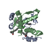
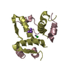
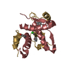
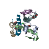
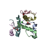
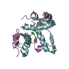
| Structure viewer | Molecule: MolmilJmol/JSmol |
|---|
- Downloads & links
-Download
| PDBx/mmCIF format | 1xw7.cif.gz | 36.3 KB | Display | PDBx/mmCIF format |
|---|---|---|---|---|
| PDB format | pdb1xw7.ent.gz | 25 KB | Display | PDB format |
| PDBx/mmJSON format | 1xw7.json.gz | Tree view | PDBx/mmJSON format | |
| Others |  Other downloads Other downloads |
-Validation report
| Arichive directory | https://data.pdbj.org/pub/pdb/validation_reports/xw/1xw7ftp://data.pdbj.org/pub/pdb/validation_reports/xw/1xw7 | HTTPS FTP |
|---|
-Related structure data
| Related structure data | 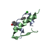 1zegS S: Starting model for refinement |
|---|---|
| Similar structure data |
-Links
 PDBj
PDBj


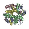
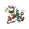
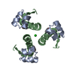
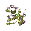
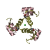
- Assembly
| Deposited unit | 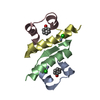
| ||||||||||||||||||
|---|---|---|---|---|---|---|---|---|---|---|---|---|---|---|---|---|---|---|---|
| 1 | 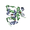
| ||||||||||||||||||
| 2 | 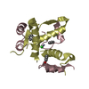
| ||||||||||||||||||
| 3 | 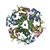
| ||||||||||||||||||
| 4 | 
| ||||||||||||||||||
| Unit cell |
| ||||||||||||||||||
| Components on special symmetry positions |
| ||||||||||||||||||
| Details | The biological assembly is a hexamer generated from the dimer in the asymmetric unit by the operations |
-Components
-Protein/peptide , 2 types, 4 molecules ACBD
| #1: Protein/peptide | Mass: 2397.725 Da / Num. of mol.: 2 / Mutation: V3L / Source method: obtained synthetically Details: The peptide was chemically synthesized. The sequence of the peptide is naturally found in Homo sapiens (human). References: UniProt: P01308 #2: Protein/peptide | Mass: 3433.953 Da / Num. of mol.: 2 / Source method: obtained synthetically Details: The peptide was chemically synthesized. The sequence of the peptide is naturally found in Homo sapiens (human). References: UniProt: P01308 |
|---|
-Non-polymers , 4 types, 86 molecules 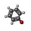


| #3: Chemical |  Mass: 94.111 Da / Num. of mol.: 2 / Source method: obtained synthetically / Formula: C6H6O Mass: 94.111 Da / Num. of mol.: 2 / Source method: obtained synthetically / Formula: C6H6O#4: Chemical |  Mass: 65.409 Da / Num. of mol.: 2 / Source method: obtained synthetically / Formula: Zn Mass: 65.409 Da / Num. of mol.: 2 / Source method: obtained synthetically / Formula: Zn#5: Chemical |  Mass: 35.453 Da / Num. of mol.: 2 / Source method: obtained synthetically / Formula: Cl Mass: 35.453 Da / Num. of mol.: 2 / Source method: obtained synthetically / Formula: Cl#6: Water | ChemComp-HOH / | Mass: 18.015 Da / Num. of mol.: 80 / Source method: isolated from a natural source / Formula: H2O |
|---|
-Details
| Has protein modification | Y |
|---|
-Experimental details
-Experiment
| Experiment | Method: X-RAY DIFFRACTION / Number of used crystals: 1 |
|---|
- Sample preparation
| Crystal | Density Matthews: 2.54 Å3/Da / Density % sol: 51.6 % |
|---|---|
| Crystal grow | Temperature: 290 K / pH: 6.2 Details: Tris, sodium citrate, acetone, phenol, pH 6.2, VAPOR DIFFUSION, HANGING DROP, temperature 290K, pH 6.20 |
-Data collection
| Diffraction | Mean temperature: 100 K |
|---|---|
| Diffraction source | Source: ROTATING ANODE / Type: BRUKER / Wavelength: 1.54 |
| Detector | Type: BRUKER / Detector: CCD / Date: Jul 23, 2004 |
| Radiation | Monochromator: MIRRORS / Protocol: SINGLE WAVELENGTH / Monochromatic (M) / Laue (L): M / Scattering type: x-ray |
| Radiation wavelength | Wavelength: 1.54 Å / Relative weight: 1 |
| Reflection | Resolution: 2.3→50.1 Å / Num. obs: 5346 / % possible obs: 97.7 % / Observed criterion σ(I): 0 / Redundancy: 12.9 % / Biso Wilson estimate: 39.7 Å2 / Rmerge(I) obs: 0.065 / Net I/σ(I): 9.7 |
| Reflection shell | Resolution: 2.3→2.44 Å / Redundancy: 9.8 % / Rmerge(I) obs: 0.066 / Mean I/σ(I) obs: 9.6 / % possible all: 95.2 |
- Processing
| Software |
| ||||||||||||||||||||||||||||||||||||||||||||||||||||||||||||||||||||||||||||||||
|---|---|---|---|---|---|---|---|---|---|---|---|---|---|---|---|---|---|---|---|---|---|---|---|---|---|---|---|---|---|---|---|---|---|---|---|---|---|---|---|---|---|---|---|---|---|---|---|---|---|---|---|---|---|---|---|---|---|---|---|---|---|---|---|---|---|---|---|---|---|---|---|---|---|---|---|---|---|---|---|---|---|
| Refinement | Method to determine structure: MOLECULAR REPLACEMENT Starting model: PDB ENTRY 1ZEG Resolution: 2.3→50.1 Å / Rfactor Rfree error: 0.009 / Isotropic thermal model: RESTRAINED / Cross valid method: THROUGHOUT / σ(F): 0 / Stereochemistry target values: ENGH & HUBER
| ||||||||||||||||||||||||||||||||||||||||||||||||||||||||||||||||||||||||||||||||
| Solvent computation | Solvent model: FLAT MODEL / Bsol: 71.24 Å2 / ksol: 0.38 e/Å3 | ||||||||||||||||||||||||||||||||||||||||||||||||||||||||||||||||||||||||||||||||
| Displacement parameters | Biso mean: 46.9 Å2
| ||||||||||||||||||||||||||||||||||||||||||||||||||||||||||||||||||||||||||||||||
| Refine analyze |
| ||||||||||||||||||||||||||||||||||||||||||||||||||||||||||||||||||||||||||||||||
| Refinement step | Cycle: LAST / Resolution: 2.3→50.1 Å
| ||||||||||||||||||||||||||||||||||||||||||||||||||||||||||||||||||||||||||||||||
| Refine LS restraints |
| ||||||||||||||||||||||||||||||||||||||||||||||||||||||||||||||||||||||||||||||||
| LS refinement shell | Resolution: 2.3→2.44 Å / Rfactor Rfree error: 0.013 / Total num. of bins used: 6
|
