 Movie
Movie Controller

[English] 日本語
 Yorodumi
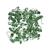
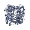
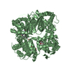
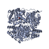
- PDB-6eds: Structure of Cysteine-free Human Insulin-Degrading Enzyme in comp... -
+ Open data

- Basic information
| Entry | Database: PDB / ID: 6eds | ||||||
|---|---|---|---|---|---|---|---|
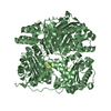
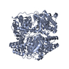
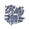
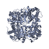
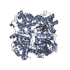
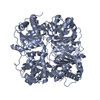
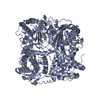
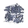
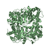
| Title | Structure of Cysteine-free Human Insulin-Degrading Enzyme in complex with Glucagon and Substrate-selective Macrocyclic Inhibitor 63 | ||||||
 Components Components |
| ||||||
 Keywords Keywords | HYDROLASE/INHIBITOR / Insulin / Glucagon / Diabetes / Exo-site / HYDROLASE / HYDROLASE-INHIBITOR complex | ||||||
| Function / homology |  Function and homology information Function and homology informationglucagon receptor binding / insulysin / beta-endorphin binding / ubiquitin recycling / insulin catabolic process / insulin metabolic process / amyloid-beta clearance by cellular catabolic process / : / hormone catabolic process / bradykinin catabolic process ...glucagon receptor binding / insulysin / beta-endorphin binding / ubiquitin recycling / insulin catabolic process / insulin metabolic process / amyloid-beta clearance by cellular catabolic process / : / hormone catabolic process / bradykinin catabolic process / feeding behavior / negative regulation of execution phase of apoptosis / cytosolic proteasome complex / positive regulation of protein binding / insulin binding / regulation of aerobic respiration / peptide catabolic process / positive regulation of calcium ion import / regulation of insulin secretion / peroxisomal matrix / amyloid-beta clearance / Synthesis, secretion, and deacylation of Ghrelin / amyloid-beta metabolic process / Insulin receptor recycling / negative regulation of proteolysis / peptide binding / cellular response to glucagon stimulus / positive regulation of gluconeogenesis / : / positive regulation of insulin secretion involved in cellular response to glucose stimulus / response to activity / gluconeogenesis / protein catabolic process / Peroxisomal protein import / hormone activity / antigen processing and presentation of endogenous peptide antigen via MHC class I / metalloendopeptidase activity / adenylate cyclase-modulating G protein-coupled receptor signaling pathway / positive regulation of insulin secretion / positive regulation of protein catabolic process / insulin receptor signaling pathway / Glucagon signaling in metabolic regulation / Synthesis, secretion, and inactivation of Glucagon-like Peptide-1 (GLP-1) / Glucagon-type ligand receptors / Glucagon-like Peptide-1 (GLP1) regulates insulin secretion / peroxisome / glucose homeostasis / amyloid-beta binding / adenylate cyclase-activating G protein-coupled receptor signaling pathway / virus receptor activity / secretory granule lumen / endopeptidase activity / G alpha (s) signalling events / basolateral plasma membrane / G alpha (q) signalling events / positive regulation of ERK1 and ERK2 cascade / Ub-specific processing proteases / endoplasmic reticulum lumen / G protein-coupled receptor signaling pathway / receptor ligand activity / signaling receptor binding / external side of plasma membrane / negative regulation of apoptotic process / protein-containing complex binding / cell surface / protein homodimerization activity / ATP hydrolysis activity / mitochondrion / proteolysis / : / extracellular exosome / extracellular region / zinc ion binding / ATP binding / identical protein binding / nucleus / plasma membrane / cytoplasm / cytosol Similarity search - Function | ||||||
| Biological species |  Homo sapiens (human) Homo sapiens (human) | ||||||
| Method |  X-RAY DIFFRACTION / SYNCHROTRON / MOLECULAR REPLACEMENT / Resolution: 3.18071730876 Å X-RAY DIFFRACTION / SYNCHROTRON / MOLECULAR REPLACEMENT / Resolution: 3.18071730876 Å | ||||||
 Authors Authors | Tan, G.A. / Seeliger, M.A. / Maianti, J.P. / Liu, D.R. / Welsh, A.J. | ||||||
 Citation Citation | Journal: Nat.Chem.Biol. / Year: 2019 Title: Substrate-selective inhibitors that reprogram the activity of insulin-degrading enzyme. Authors: Maianti, J.P. / Tan, G.A. / Vetere, A. / Welsh, A.J. / Wagner, B.K. / Seeliger, M.A. / Liu, D.R. #1: Journal: Nature / Year: 2014Title: Anti-diabetic activity of insulin-degrading enzyme inhibitors mediated by multiple hormones. Authors: Maianti, J.P. / McFedries, A. / Foda, Z.H. / Kleiner, R.E. / Du, X.Q. / Leissring, M.A. / Tang, W.J. / Charron, M.J. / Seeliger, M.A. / Saghatelian, A. / Liu, D.R. | ||||||
| History |
|
- Structure visualization
| Structure viewer | Molecule: MolmilJmol/JSmol |
|---|
- Downloads & links
-Download
| PDBx/mmCIF format | 6eds.cif.gz | 954 KB | Display | PDBx/mmCIF format |
|---|---|---|---|---|
| PDB format | pdb6eds.ent.gz | 648.1 KB | Display | PDB format |
| PDBx/mmJSON format | 6eds.json.gz | Tree view | PDBx/mmJSON format | |
| Others |  Other downloads Other downloads |
-Validation report
| Arichive directory | https://data.pdbj.org/pub/pdb/validation_reports/ed/6edsftp://data.pdbj.org/pub/pdb/validation_reports/ed/6eds | HTTPS FTP |
|---|
-Related structure data
| Related structure data |  6byzC  6mq3C  4lteS S: Starting model for refinement C: citing same article ( |
|---|---|
| Similar structure data |


-Links
 PDBj
PDBj


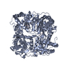
- Assembly
| Deposited unit | 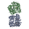
| |||||||||||||||||||||||||||||||||||||||||||||||||||||||||||||||||||||||||||||||||||||||||||||||||||||||||||||||||||||||||
|---|---|---|---|---|---|---|---|---|---|---|---|---|---|---|---|---|---|---|---|---|---|---|---|---|---|---|---|---|---|---|---|---|---|---|---|---|---|---|---|---|---|---|---|---|---|---|---|---|---|---|---|---|---|---|---|---|---|---|---|---|---|---|---|---|---|---|---|---|---|---|---|---|---|---|---|---|---|---|---|---|---|---|---|---|---|---|---|---|---|---|---|---|---|---|---|---|---|---|---|---|---|---|---|---|---|---|---|---|---|---|---|---|---|---|---|---|---|---|---|---|---|---|
| 1 | 
| |||||||||||||||||||||||||||||||||||||||||||||||||||||||||||||||||||||||||||||||||||||||||||||||||||||||||||||||||||||||||
| 2 | 
| |||||||||||||||||||||||||||||||||||||||||||||||||||||||||||||||||||||||||||||||||||||||||||||||||||||||||||||||||||||||||
| Unit cell |
| |||||||||||||||||||||||||||||||||||||||||||||||||||||||||||||||||||||||||||||||||||||||||||||||||||||||||||||||||||||||||
| Noncrystallographic symmetry (NCS) | NCS domain:
NCS domain segments:
NCS ensembles :
|
-Components
-Protein / Protein/peptide , 2 types, 4 molecules ABCD
| #1: Protein | Mass: 113191.031 Da / Num. of mol.: 2 Mutation: C110L, E111Q, C171S, C178A, C257V, C414L, C573N, C590S, C789S, C812A, C819A, C904S, C966N, C974A Source method: isolated from a genetically manipulated source Source: (gene. exp.) Homo sapiens (human) / Gene: IDE / Production host:  #2: Protein/peptide | Mass: 3486.781 Da / Num. of mol.: 2 Source method: isolated from a genetically manipulated source Details: Glucagon, marketed as Glucagen and manufactured by Boehringer Ingelheim. Source: (gene. exp.) Homo sapiens (human) / Gene: GCG / Production host:  |
|---|
-Non-polymers , 4 types, 8 molecules 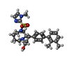


| #3: Chemical |  Mass: 494.649 Da / Num. of mol.: 2 / Source method: obtained synthetically / Formula: C27H34N4O3S / Feature type: SUBJECT OF INVESTIGATION Mass: 494.649 Da / Num. of mol.: 2 / Source method: obtained synthetically / Formula: C27H34N4O3S / Feature type: SUBJECT OF INVESTIGATION#4: Chemical |  Mass: 238.305 Da / Num. of mol.: 2 / Source method: obtained synthetically / Formula: C8H18N2O4S / Comment: pH buffer*YM Mass: 238.305 Da / Num. of mol.: 2 / Source method: obtained synthetically / Formula: C8H18N2O4S / Comment: pH buffer*YM#5: Chemical |  Mass: 106.120 Da / Num. of mol.: 2 / Source method: obtained synthetically / Formula: C4H10O3 Mass: 106.120 Da / Num. of mol.: 2 / Source method: obtained synthetically / Formula: C4H10O3#6: Chemical |  Mass: 88.105 Da / Num. of mol.: 2 / Source method: obtained synthetically / Formula: C4H8O2 Mass: 88.105 Da / Num. of mol.: 2 / Source method: obtained synthetically / Formula: C4H8O2 |
|---|
-Experimental details
-Experiment
| Experiment | Method: X-RAY DIFFRACTION / Number of used crystals: 1 |
|---|
- Sample preparation
| Crystal | Density Matthews: 4.01 Å3/Da / Density % sol: 69.32 % |
|---|---|
| Crystal grow | Temperature: 298 K / Method: vapor diffusion, hanging drop Details: 0.1M HEPES pH 7.0, 12% Tacsimate pH 7.0, 13% PEGMME, 10% Dioxane PH range: 6.8-7.0 / Temp details: Room temperature |
-Data collection
| Diffraction | Mean temperature: 100 K |
|---|---|
| Diffraction source | Source: SYNCHROTRON / Site: NSLS-II  / Beamline: 17-ID-2 / Wavelength: 0.979341 Å / Beamline: 17-ID-2 / Wavelength: 0.979341 Å |
| Detector | Type: DECTRIS EIGER X 16M / Detector: PIXEL / Date: Jul 19, 2018 |
| Radiation | Protocol: SINGLE WAVELENGTH / Monochromatic (M) / Laue (L): M / Scattering type: x-ray |
| Radiation wavelength | Wavelength: 0.979341 Å / Relative weight: 1 |
| Reflection | Resolution: 3.18→131.559 Å / Num. obs: 60302 / % possible obs: 100 % / Redundancy: 20.9 % / Biso Wilson estimate: 57.9001204686 Å2 / CC1/2: 0.991 / Rmerge(I) obs: 0.369 / Rpim(I) all: 0.083 / Rrim(I) all: 0.378 / Net I/σ(I): 10.98 |
| Reflection shell | Resolution: 3.181→3.2356 Å |
- Processing
| Software |
| |||||||||||||||||||||||||||||||||||||||||||||||||||||||||||||||||||||||||||||||||||||||||||||||||||||||||||||||||||||||||||||||||||||||||||||||||||
|---|---|---|---|---|---|---|---|---|---|---|---|---|---|---|---|---|---|---|---|---|---|---|---|---|---|---|---|---|---|---|---|---|---|---|---|---|---|---|---|---|---|---|---|---|---|---|---|---|---|---|---|---|---|---|---|---|---|---|---|---|---|---|---|---|---|---|---|---|---|---|---|---|---|---|---|---|---|---|---|---|---|---|---|---|---|---|---|---|---|---|---|---|---|---|---|---|---|---|---|---|---|---|---|---|---|---|---|---|---|---|---|---|---|---|---|---|---|---|---|---|---|---|---|---|---|---|---|---|---|---|---|---|---|---|---|---|---|---|---|---|---|---|---|---|---|---|---|---|
| Refinement | Method to determine structure: MOLECULAR REPLACEMENT Starting model: 4LTE Resolution: 3.18071730876→131.559 Å / SU ML: 0.332870484009 / Cross valid method: FREE R-VALUE / σ(F): 1.33583552402 / Phase error: 21.3338903277
| |||||||||||||||||||||||||||||||||||||||||||||||||||||||||||||||||||||||||||||||||||||||||||||||||||||||||||||||||||||||||||||||||||||||||||||||||||
| Solvent computation | Shrinkage radii: 0.9 Å / VDW probe radii: 1.11 Å | |||||||||||||||||||||||||||||||||||||||||||||||||||||||||||||||||||||||||||||||||||||||||||||||||||||||||||||||||||||||||||||||||||||||||||||||||||
| Displacement parameters | Biso mean: 51.5855318537 Å2 | |||||||||||||||||||||||||||||||||||||||||||||||||||||||||||||||||||||||||||||||||||||||||||||||||||||||||||||||||||||||||||||||||||||||||||||||||||
| Refinement step | Cycle: LAST / Resolution: 3.18071730876→131.559 Å
| |||||||||||||||||||||||||||||||||||||||||||||||||||||||||||||||||||||||||||||||||||||||||||||||||||||||||||||||||||||||||||||||||||||||||||||||||||
| Refine LS restraints |
| |||||||||||||||||||||||||||||||||||||||||||||||||||||||||||||||||||||||||||||||||||||||||||||||||||||||||||||||||||||||||||||||||||||||||||||||||||
| LS refinement shell |
| |||||||||||||||||||||||||||||||||||||||||||||||||||||||||||||||||||||||||||||||||||||||||||||||||||||||||||||||||||||||||||||||||||||||||||||||||||
| Refinement TLS params. | Method: refined / Origin x: 72.4902556943 Å / Origin y: -78.3115153621 Å / Origin z: 4.86863644608 Å
| |||||||||||||||||||||||||||||||||||||||||||||||||||||||||||||||||||||||||||||||||||||||||||||||||||||||||||||||||||||||||||||||||||||||||||||||||||
| Refinement TLS group | Selection details: all |
