 Movie
Movie Controller

[English] 日本語
 Yorodumi
- PDB-4ral: Crystal structure of insulin degrading enzyme in complex with mac... -
+ Open data

- Basic information
| Entry | Database: PDB / ID: 4ral | |||||||||
|---|---|---|---|---|---|---|---|---|---|---|
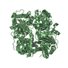
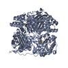
| Title | Crystal structure of insulin degrading enzyme in complex with macrophage inflammatory protein 1 beta | |||||||||
 Components Components |
| |||||||||
 Keywords Keywords | HYDROLASE/CYTOKINE / IDE / MIP1alpha / metal-binding / metalloprotease / chemotaxis / inflammatory response / HYDROLASE-CYTOKINE complex | |||||||||
| Function / homology |  Function and homology information Function and homology informationCCR1 chemokine receptor binding / positive regulation of natural killer cell chemotaxis / insulysin / beta-endorphin binding / ubiquitin recycling / insulin catabolic process / CCR5 chemokine receptor binding / CCR chemokine receptor binding / insulin metabolic process / amyloid-beta clearance by cellular catabolic process ...CCR1 chemokine receptor binding / positive regulation of natural killer cell chemotaxis / insulysin / beta-endorphin binding / ubiquitin recycling / insulin catabolic process / CCR5 chemokine receptor binding / CCR chemokine receptor binding / insulin metabolic process / amyloid-beta clearance by cellular catabolic process / hormone catabolic process / bradykinin catabolic process / eosinophil chemotaxis / positive regulation of calcium ion transport / cytosolic proteasome complex / chemokine activity / positive regulation of protein binding / Chemokine receptors bind chemokines / insulin binding / regulation of aerobic respiration / peptide catabolic process / Interleukin-10 signaling / peroxisomal matrix / establishment or maintenance of cell polarity / amyloid-beta clearance / host-mediated suppression of viral transcription / amyloid-beta metabolic process / Insulin receptor recycling / negative regulation of proteolysis / peptide binding / positive regulation of calcium-mediated signaling / : / cytokine activity / chemokine-mediated signaling pathway / protein catabolic process / Peroxisomal protein import / antigen processing and presentation of endogenous peptide antigen via MHC class I / metalloendopeptidase activity / response to toxic substance / response to virus / positive regulation of protein catabolic process / insulin receptor signaling pathway / peroxisome / cell-cell signaling / antimicrobial humoral immune response mediated by antimicrobial peptide / amyloid-beta binding / virus receptor activity / endopeptidase activity / G alpha (i) signalling events / basolateral plasma membrane / cell adhesion / Ub-specific processing proteases / immune response / positive regulation of cell migration / inflammatory response / external side of plasma membrane / protein-containing complex binding / cell surface / signal transduction / protein homodimerization activity / ATP hydrolysis activity / mitochondrion / proteolysis / : / extracellular exosome / extracellular region / zinc ion binding / ATP binding / identical protein binding / nucleus / cytoplasm / cytosol Similarity search - Function | |||||||||
| Biological species |  Homo sapiens (human) Homo sapiens (human) | |||||||||
| Method |  X-RAY DIFFRACTION / SYNCHROTRON / MOLECULAR REPLACEMENT / Resolution: 3.148 Å X-RAY DIFFRACTION / SYNCHROTRON / MOLECULAR REPLACEMENT / Resolution: 3.148 Å | |||||||||
 Authors Authors | Liang, W.G. / Ren, M. / Guo, Q. / Tang, W.J. | |||||||||
 Citation Citation | Journal: J.Mol.Biol. / Year: 2015 Title: Structures of human CCL18, CCL3, and CCL4 reveal molecular determinants for quaternary structures and sensitivity to insulin-degrading enzyme. Authors: Liang, W.G. / Ren, M. / Zhao, F. / Tang, W.J. | |||||||||
| History |
|
- Structure visualization
| Structure viewer | Molecule: MolmilJmol/JSmol |
|---|
- Downloads & links
-Download
| PDBx/mmCIF format | 4ral.cif.gz | 788.5 KB | Display | PDBx/mmCIF format |
|---|---|---|---|---|
| PDB format | pdb4ral.ent.gz | 647 KB | Display | PDB format |
| PDBx/mmJSON format | 4ral.json.gz | Tree view | PDBx/mmJSON format | |
| Others |  Other downloads Other downloads |
-Validation report
| Arichive directory | https://data.pdbj.org/pub/pdb/validation_reports/ra/4ralftp://data.pdbj.org/pub/pdb/validation_reports/ra/4ral | HTTPS FTP |
|---|
-Related structure data
| Related structure data | 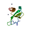 3tn2C 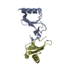 4mheC  4ra8C  3cwwS S: Starting model for refinement C: citing same article ( |
|---|---|
| Similar structure data |


-Links
 PDBj
PDBj


- Assembly
| Deposited unit | 
| ||||||||
|---|---|---|---|---|---|---|---|---|---|
| 1 | 
| ||||||||
| 2 | 
| ||||||||
| Unit cell |
|
-Components
| #1: Protein | Mass: 114560.578 Da / Num. of mol.: 2 / Fragment: UNP residues 42-1019 Mutation: C110L, E111Q, C171S, C178A, C257V, C414L, C573N, C590S, C789S, C812A, C819A, C904S, C966N, C974A Source method: isolated from a genetically manipulated source Source: (gene. exp.) Homo sapiens (human) / Gene: IDE / Production host:  #2: Protein | Mass: 7824.742 Da / Num. of mol.: 2 / Fragment: UNP residues 24-92 Source method: isolated from a genetically manipulated source Source: (gene. exp.) Homo sapiens (human) / Gene: CCL4, LAG1, MIP1B, SCYA4 / Production host: #3: Chemical |   Mass: 65.409 Da / Num. of mol.: 2 / Source method: obtained synthetically / Formula: Zn Mass: 65.409 Da / Num. of mol.: 2 / Source method: obtained synthetically / Formula: Zn#4: Water | ChemComp-HOH / |  Mass: 18.015 Da / Num. of mol.: 67 / Source method: isolated from a natural source / Formula: H2O Mass: 18.015 Da / Num. of mol.: 67 / Source method: isolated from a natural source / Formula: H2O |
|---|
-Experimental details
-Experiment
| Experiment | Method: X-RAY DIFFRACTION / Number of used crystals: 1 |
|---|
- Sample preparation
| Crystal | Density Matthews: 3.71 Å3/Da / Density % sol: 66.84 % |
|---|---|
| Crystal grow | Temperature: 291 K / Method: vapor diffusion, hanging drop / pH: 7 Details: 13% PEG5000 MME, 100 mM HEPES, pH 7.0, 10% Tacsimate, 10% dioxane, VAPOR DIFFUSION, HANGING DROP, temperature 291.0K |
-Data collection
| Diffraction | Mean temperature: 100 K |
|---|---|
| Diffraction source | Source: SYNCHROTRON / Site: APS  / Beamline: 19-ID / Beamline: 19-ID |
| Detector | Type: ADSC QUANTUM 315r / Detector: CCD / Date: Jul 8, 2010 |
| Radiation | Monochromator: Rosenbaum-Rock high-resolution double-crystal Si(111) Protocol: SINGLE WAVELENGTH / Monochromatic (M) / Laue (L): M / Scattering type: x-ray |
| Radiation wavelength | Relative weight: 1 |
| Reflection | Resolution: 3.148→48.263 Å / Num. all: 59484 / Num. obs: 59484 / % possible obs: 95.5 % / Observed criterion σ(F): 3.06 / Observed criterion σ(I): 3.06 / Redundancy: 4.3 % / Rmerge(I) obs: 0.209 / Rsym value: 0.216 / Net I/σ(I): 7.4 |
| Reflection shell | Highest resolution: 3.148 Å |
- Processing
| Software |
| |||||||||||||||||||||||||||||||||||||||||||||||||
|---|---|---|---|---|---|---|---|---|---|---|---|---|---|---|---|---|---|---|---|---|---|---|---|---|---|---|---|---|---|---|---|---|---|---|---|---|---|---|---|---|---|---|---|---|---|---|---|---|---|---|
| Refinement | Method to determine structure: MOLECULAR REPLACEMENT Starting model: PDB ENTRY 3CWW Resolution: 3.148→48.263 Å / SU ML: 0.31 / σ(F): 1.34 / Phase error: 25.9 / Stereochemistry target values: ML
| |||||||||||||||||||||||||||||||||||||||||||||||||
| Solvent computation | Shrinkage radii: 0.9 Å / VDW probe radii: 1.11 Å / Solvent model: FLAT BULK SOLVENT MODEL | |||||||||||||||||||||||||||||||||||||||||||||||||
| Refinement step | Cycle: LAST / Resolution: 3.148→48.263 Å
| |||||||||||||||||||||||||||||||||||||||||||||||||
| Refine LS restraints |
| |||||||||||||||||||||||||||||||||||||||||||||||||
| LS refinement shell |
| |||||||||||||||||||||||||||||||||||||||||||||||||
| Refinement TLS params. | Method: refined / Origin x: -104.0282 Å / Origin y: 23.6136 Å / Origin z: -5.9716 Å
| |||||||||||||||||||||||||||||||||||||||||||||||||
| Refinement TLS group | Selection details: all |
