 Movie
Movie Controller

[English] 日本語
 Yorodumi
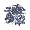
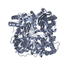
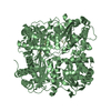
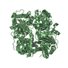
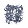
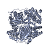
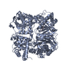
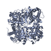
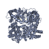
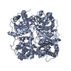
- PDB-2g56: crystal structure of human insulin-degrading enzyme in complex wi... -
+ Open data

- Basic information
| Entry | Database: PDB / ID: 2g56 | ||||||
|---|---|---|---|---|---|---|---|
| Title | crystal structure of human insulin-degrading enzyme in complex with insulin B chain | ||||||
 Components Components |
| ||||||
 Keywords Keywords | HYDROLASE / protein-peptide complex | ||||||
| Function / homology |  Function and homology information Function and homology informationinsulysin / beta-endorphin binding / ubiquitin recycling / insulin catabolic process / insulin metabolic process / amyloid-beta clearance by cellular catabolic process / hormone catabolic process / bradykinin catabolic process / cytosolic proteasome complex / positive regulation of protein binding ...insulysin / beta-endorphin binding / ubiquitin recycling / insulin catabolic process / insulin metabolic process / amyloid-beta clearance by cellular catabolic process / hormone catabolic process / bradykinin catabolic process / cytosolic proteasome complex / positive regulation of protein binding / insulin binding / negative regulation of glycogen catabolic process / : / regulation of aerobic respiration / negative regulation of fatty acid metabolic process / Signaling by Insulin receptor / negative regulation of feeding behavior / IRS activation / Insulin processing / peptide catabolic process / regulation of protein secretion / positive regulation of peptide hormone secretion / negative regulation of acute inflammatory response / Regulation of gene expression in beta cells / positive regulation of respiratory burst / peroxisomal matrix / alpha-beta T cell activation / amyloid-beta clearance / Synthesis, secretion, and deacylation of Ghrelin / amyloid-beta metabolic process / negative regulation of protein secretion / positive regulation of dendritic spine maintenance / negative regulation of gluconeogenesis / fatty acid homeostasis / positive regulation of glycogen biosynthetic process / positive regulation of insulin receptor signaling pathway / Signal attenuation / FOXO-mediated transcription of oxidative stress, metabolic and neuronal genes / negative regulation of lipid catabolic process / negative regulation of respiratory burst involved in inflammatory response / positive regulation of lipid biosynthetic process / negative regulation of oxidative stress-induced intrinsic apoptotic signaling pathway / nitric oxide-cGMP-mediated signaling / regulation of protein localization to plasma membrane / positive regulation of nitric-oxide synthase activity / transport vesicle / Insulin receptor recycling / COPI-mediated anterograde transport / negative regulation of reactive oxygen species biosynthetic process / positive regulation of brown fat cell differentiation / insulin-like growth factor receptor binding / negative regulation of proteolysis / NPAS4 regulates expression of target genes / peptide binding / neuron projection maintenance / positive regulation of mitotic nuclear division / endoplasmic reticulum-Golgi intermediate compartment membrane / Insulin receptor signalling cascade / : / positive regulation of glycolytic process / endosome lumen / positive regulation of cytokine production / acute-phase response / positive regulation of D-glucose import across plasma membrane / insulin receptor binding / protein catabolic process / positive regulation of long-term synaptic potentiation / positive regulation of protein secretion / positive regulation of cell differentiation / wound healing / Peroxisomal protein import / Regulation of insulin secretion / hormone activity / antigen processing and presentation of endogenous peptide antigen via MHC class I / positive regulation of neuron projection development / metalloendopeptidase activity / negative regulation of protein catabolic process / regulation of synaptic plasticity / positive regulation of protein localization to nucleus / Golgi lumen / cognition / glucose metabolic process / vasodilation / positive regulation of protein catabolic process / insulin receptor signaling pathway / cell-cell signaling / peroxisome / regulation of protein localization / glucose homeostasis / amyloid-beta binding / PI5P, PP2A and IER3 Regulate PI3K/AKT Signaling / virus receptor activity / positive regulation of cell growth / protease binding / secretory granule lumen / endopeptidase activity / basolateral plasma membrane / positive regulation of MAPK cascade / positive regulation of canonical NF-kappaB signal transduction / positive regulation of phosphatidylinositol 3-kinase/protein kinase B signal transduction Similarity search - Function | ||||||
| Biological species |  Homo sapiens (human) Homo sapiens (human) | ||||||
| Method |  X-RAY DIFFRACTION / SYNCHROTRON / MOLECULAR REPLACEMENT / Resolution: 2.2 Å X-RAY DIFFRACTION / SYNCHROTRON / MOLECULAR REPLACEMENT / Resolution: 2.2 Å | ||||||
 Authors Authors | Shen, Y. / Tang, W.-J. | ||||||
 Citation Citation | Journal: Nature / Year: 2006 Title: Structures of human insulin-degrading enzyme reveal a new substrate recognition mechanism. Authors: Shen, Y. / Joachimiak, A. / Rosner, M.R. / Tang, W.J. | ||||||
| History |
|
- Structure visualization
| Structure viewer | Molecule: MolmilJmol/JSmol |
|---|
- Downloads & links
-Download
| PDBx/mmCIF format | 2g56.cif.gz | 431.3 KB | Display | PDBx/mmCIF format |
|---|---|---|---|---|
| PDB format | pdb2g56.ent.gz | 338.9 KB | Display | PDB format |
| PDBx/mmJSON format | 2g56.json.gz | Tree view | PDBx/mmJSON format | |
| Others |  Other downloads Other downloads |
-Validation report
| Arichive directory | https://data.pdbj.org/pub/pdb/validation_reports/g5/2g56ftp://data.pdbj.org/pub/pdb/validation_reports/g5/2g56 | HTTPS FTP |
|---|
-Related structure data
| Related structure data |  2g47C  2g48C  2g49C  2g54SC C: citing same article ( S: Starting model for refinement |
|---|---|
| Similar structure data |


-Links
 PDBj
PDBj


- Assembly
| Deposited unit | 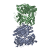
| ||||||||
|---|---|---|---|---|---|---|---|---|---|
| 1 | 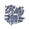
| ||||||||
| 2 | 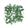
| ||||||||
| Unit cell |
| ||||||||
| Details | the biological assembly is a monomer. |
-Components
| #1: Protein | Mass: 114715.141 Da / Num. of mol.: 2 Source method: isolated from a genetically manipulated source Source: (gene. exp.) Homo sapiens (human) / Gene: IDE / Plasmid: pProEx / Production host:  References: UniProt: Q5T5N2, UniProt: P14735*PLUS, insulysin #2: Protein/peptide | Mass: 3433.953 Da / Num. of mol.: 2 / Fragment: Insulin B chain, residues 25-54 / Source method: obtained synthetically Details: This sequence occurs naturally in Homo sapiens (Humans) References: UniProt: P01308 #3: Chemical | ChemComp-DIO / |   Mass: 88.105 Da / Num. of mol.: 1 / Source method: obtained synthetically / Formula: C4H8O2 Mass: 88.105 Da / Num. of mol.: 1 / Source method: obtained synthetically / Formula: C4H8O2#4: Water | ChemComp-HOH / |  Mass: 18.015 Da / Num. of mol.: 1044 / Source method: isolated from a natural source / Formula: H2O Mass: 18.015 Da / Num. of mol.: 1044 / Source method: isolated from a natural source / Formula: H2O |
|---|
-Experimental details
-Experiment
| Experiment | Method: X-RAY DIFFRACTION / Number of used crystals: 1 |
|---|
- Sample preparation
| Crystal | Density Matthews: 3.8 Å3/Da / Density % sol: 67.67 % |
|---|---|
| Crystal grow | Temperature: 298 K / Method: vapor diffusion, hanging drop / pH: 7 Details: PEGMME5000, dioxane, tacismate, hepes, pH 7.0, VAPOR DIFFUSION, HANGING DROP, temperature 298K |
-Data collection
| Diffraction | Mean temperature: 100 K |
|---|---|
| Diffraction source | Source: SYNCHROTRON / Site: APS  / Beamline: 19-ID / Wavelength: 0.9 Å / Beamline: 19-ID / Wavelength: 0.9 Å |
| Detector | Type: ADSC QUANTUM 4 / Detector: CCD / Date: Aug 23, 2005 |
| Radiation | Monochromator: graphite / Protocol: SINGLE WAVELENGTH / Monochromatic (M) / Laue (L): M / Scattering type: x-ray |
| Radiation wavelength | Wavelength: 0.9 Å / Relative weight: 1 |
| Reflection | Resolution: 2.2→30 Å / Num. all: 175528 / Num. obs: 175528 / % possible obs: 99 % / Observed criterion σ(F): 2 / Observed criterion σ(I): 2 / Redundancy: 8.4 % / Biso Wilson estimate: 20.7 Å2 / Rsym value: 0.064 / Net I/σ(I): 26.5 |
| Reflection shell | Resolution: 2.2→2.28 Å / Redundancy: 5.7 % / Mean I/σ(I) obs: 4.6 / Num. unique all: 15806 / Rsym value: 0.347 / % possible all: 87.9 |
- Processing
| Software |
| ||||||||||||||||||||||||||||||||||||
|---|---|---|---|---|---|---|---|---|---|---|---|---|---|---|---|---|---|---|---|---|---|---|---|---|---|---|---|---|---|---|---|---|---|---|---|---|---|
| Refinement | Method to determine structure: MOLECULAR REPLACEMENT Starting model: PDB ENTRY 2G54 Resolution: 2.2→29.75 Å / Rfactor Rfree error: 0.002 / Data cutoff high absF: 142221.1 / Data cutoff low absF: 0 / Isotropic thermal model: RESTRAINED / Cross valid method: THROUGHOUT / σ(F): 0
| ||||||||||||||||||||||||||||||||||||
| Solvent computation | Solvent model: FLAT MODEL / Bsol: 51.0152 Å2 / ksol: 0.3739 e/Å3 | ||||||||||||||||||||||||||||||||||||
| Displacement parameters | Biso mean: 32.2 Å2
| ||||||||||||||||||||||||||||||||||||
| Refine analyze |
| ||||||||||||||||||||||||||||||||||||
| Refinement step | Cycle: LAST / Resolution: 2.2→29.75 Å
| ||||||||||||||||||||||||||||||||||||
| Refine LS restraints |
| ||||||||||||||||||||||||||||||||||||
| LS refinement shell | Resolution: 2.2→2.34 Å / Rfactor Rfree error: 0.005 / Total num. of bins used: 6
| ||||||||||||||||||||||||||||||||||||
| Xplor file |
|
