 Movie
Movie Controller

[English] 日本語
 Yorodumi
- PDB-3h44: Crystal Structure of Insulin Degrading Enzyme in Complex with mac... -
+ Open data

- Basic information
| Entry | Database: PDB / ID: 3h44 | ||||||
|---|---|---|---|---|---|---|---|
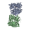
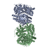
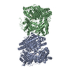
| Title | Crystal Structure of Insulin Degrading Enzyme in Complex with macrophage inflammatory protein 1 alpha | ||||||
 Components Components |
| ||||||
 Keywords Keywords | Hydrolase/Cytokine / IDE / MIP1alpha / Cytoplasm / Hydrolase / Metal-binding / Metalloprotease / Polymorphism / Protease / Zinc / Chemotaxis / Cytokine / Disulfide bond / Inflammatory response / Secreted / Hydrolase-Cytokine COMPLEX | ||||||
| Function / homology |  Function and homology information Function and homology informationlymphocyte chemotaxis / granulocyte chemotaxis / CCR1 chemokine receptor binding / positive regulation of natural killer cell chemotaxis / positive regulation of microglial cell migration / insulysin / beta-endorphin binding / astrocyte cell migration / ubiquitin recycling / insulin catabolic process ...lymphocyte chemotaxis / granulocyte chemotaxis / CCR1 chemokine receptor binding / positive regulation of natural killer cell chemotaxis / positive regulation of microglial cell migration / insulysin / beta-endorphin binding / astrocyte cell migration / ubiquitin recycling / insulin catabolic process / regulation of behavior / CCR5 chemokine receptor binding / eosinophil degranulation / CCR chemokine receptor binding / regulation of sensory perception of pain / insulin metabolic process / signaling / positive regulation of microglial cell activation / amyloid-beta clearance by cellular catabolic process / cell activation / negative regulation of bone mineralization / hormone catabolic process / bradykinin catabolic process / T cell chemotaxis / eosinophil chemotaxis / positive regulation of calcium ion transport / response to cholesterol / cytosolic proteasome complex / chemokine activity / release of sequestered calcium ion into cytosol by sarcoplasmic reticulum / positive regulation of protein binding / Chemokine receptors bind chemokines / insulin binding / phospholipase activator activity / regulation of aerobic respiration / peptide catabolic process / positive regulation of calcium ion import / chemoattractant activity / negative regulation of osteoclast differentiation / Interleukin-10 signaling / exocytosis / peroxisomal matrix / macrophage chemotaxis / amyloid-beta clearance / monocyte chemotaxis / host-mediated suppression of viral transcription / amyloid-beta metabolic process / cellular response to interleukin-1 / Insulin receptor recycling / negative regulation of proteolysis / neutrophil chemotaxis / peptide binding / cytoskeleton organization / positive regulation of calcium-mediated signaling / : / positive regulation of interleukin-1 beta production / chemokine-mediated signaling pathway / protein catabolic process / cell chemotaxis / Peroxisomal protein import / calcium-mediated signaling / antigen processing and presentation of endogenous peptide antigen via MHC class I / metalloendopeptidase activity / cellular response to type II interferon / cellular response to tumor necrosis factor / response to toxic substance / chemotaxis / intracellular calcium ion homeostasis / positive regulation of tumor necrosis factor production / positive regulation of inflammatory response / kinase activity / osteoblast differentiation / positive regulation of protein catabolic process / calcium ion transport / insulin receptor signaling pathway / peroxisome / regulation of cell shape / positive regulation of neuron apoptotic process / MAPK cascade / cell-cell signaling / antimicrobial humoral immune response mediated by antimicrobial peptide / amyloid-beta binding / virus receptor activity / endopeptidase activity / basolateral plasma membrane / protein kinase activity / positive regulation of ERK1 and ERK2 cascade / positive regulation of phosphatidylinositol 3-kinase/protein kinase B signal transduction / Ub-specific processing proteases / positive regulation of cell migration / inflammatory response / external side of plasma membrane / negative regulation of gene expression / positive regulation of gene expression / protein-containing complex binding / cell surface / protein homodimerization activity / ATP hydrolysis activity / mitochondrion / proteolysis Similarity search - Function | ||||||
| Biological species |  Homo sapiens (human) Homo sapiens (human) | ||||||
| Method |  X-RAY DIFFRACTION / SYNCHROTRON / MOLECULAR REPLACEMENT / Resolution: 3 Å X-RAY DIFFRACTION / SYNCHROTRON / MOLECULAR REPLACEMENT / Resolution: 3 Å | ||||||
 Authors Authors | Ren, M. / Guo, Q. / Tang, W.J. | ||||||
 Citation Citation | Journal: To be Published Title: Macrophage Inflammatory Protein-1 Is A Novel High Affinity Substrate For Human Insulin Degrading Enzyme Authors: Ren, M. / Guo, Q. / Tang, W.J. | ||||||
| History |
|
- Structure visualization
| Structure viewer | Molecule: MolmilJmol/JSmol |
|---|
- Downloads & links
-Download
| PDBx/mmCIF format | 3h44.cif.gz | 406.6 KB | Display | PDBx/mmCIF format |
|---|---|---|---|---|
| PDB format | pdb3h44.ent.gz | 319.5 KB | Display | PDB format |
| PDBx/mmJSON format | 3h44.json.gz | Tree view | PDBx/mmJSON format | |
| Others |  Other downloads Other downloads |
-Validation report
| Arichive directory | https://data.pdbj.org/pub/pdb/validation_reports/h4/3h44ftp://data.pdbj.org/pub/pdb/validation_reports/h4/3h44 | HTTPS FTP |
|---|
-Related structure data
| Related structure data | 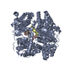 2wbyS S: Starting model for refinement |
|---|---|
| Similar structure data |

-Links
 PDBj
PDBj


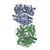
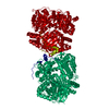
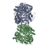
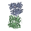
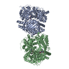
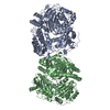
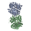
- Assembly
| Deposited unit | 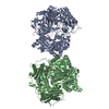
| ||||||||
|---|---|---|---|---|---|---|---|---|---|
| 1 | 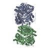
| ||||||||
| 2 |
| ||||||||
| 3 | 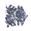
| ||||||||
| 4 | 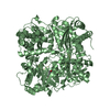
| ||||||||
| Unit cell |
|
-Components
| #1: Protein | Mass: 114560.578 Da / Num. of mol.: 2 / Fragment: residues 42-1019 Mutation: C110L, E111Q, C171S, C178A, C257V, C414L, C573N, C590S, C789S, C812A, C819A, C904S, C966N, C974A Source method: isolated from a genetically manipulated source Source: (gene. exp.) Homo sapiens (human) / Gene: IDE / Plasmid: proEX-H6 / Production host:  #2: Protein | Mass: 7793.664 Da / Num. of mol.: 2 / Fragment: residues 23-92 Source method: isolated from a genetically manipulated source Source: (gene. exp.) Homo sapiens (human) / Gene: CCL3, G0S19-1, MIP1A, MIP1alpha, SCYA3 / Plasmid: pET-32a / Production host: #3: Chemical | ChemComp-DIO /   Mass: 88.105 Da / Num. of mol.: 6 / Source method: obtained synthetically / Formula: C4H8O2 Mass: 88.105 Da / Num. of mol.: 6 / Source method: obtained synthetically / Formula: C4H8O2#4: Chemical |   Mass: 65.409 Da / Num. of mol.: 2 / Source method: obtained synthetically / Formula: Zn Mass: 65.409 Da / Num. of mol.: 2 / Source method: obtained synthetically / Formula: Zn#5: Water | ChemComp-HOH / |  Mass: 18.015 Da / Num. of mol.: 133 / Source method: isolated from a natural source / Formula: H2O Mass: 18.015 Da / Num. of mol.: 133 / Source method: isolated from a natural source / Formula: H2O |
|---|
-Experimental details
-Experiment
| Experiment | Method: X-RAY DIFFRACTION / Number of used crystals: 1 |
|---|
- Sample preparation
| Crystal | Density Matthews: 3.68 Å3/Da / Density % sol: 66.62 % |
|---|---|
| Crystal grow | Temperature: 291 K / Method: vapor diffusion, hanging drop / pH: 7 Details: 13% PEGMME 5000, 100mM HEPES, pH 7.0, 10%, Tacsimate, 10% dioxane, VAPOR DIFFUSION, HANGING DROP, temperature 291K |
-Data collection
| Diffraction | Mean temperature: 100 K |
|---|---|
| Diffraction source | Source: SYNCHROTRON / Site: APS  / Beamline: 14-BM-C / Wavelength: 0.9762 Å / Beamline: 14-BM-C / Wavelength: 0.9762 Å |
| Detector | Type: ADSC QUANTUM 315 / Detector: CCD / Date: Jan 1, 2008 / Details: bent conical Si-mirror (Rh coating) |
| Radiation | Monochromator: bent Ge(111) / Protocol: SINGLE WAVELENGTH / Monochromatic (M) / Laue (L): M / Scattering type: x-ray |
| Radiation wavelength | Wavelength: 0.9762 Å / Relative weight: 1 |
| Reflection | Resolution: 3→50 Å / Num. all: 67989 / Num. obs: 67744 / % possible obs: 99.64 % / Observed criterion σ(F): 2 / Observed criterion σ(I): 2 / Redundancy: 5 % / Rmerge(I) obs: 0.142 / Rsym value: 0.142 |
| Reflection shell | Resolution: 3→3.11 Å / Redundancy: 5 % / Rmerge(I) obs: 0.53 / Mean I/σ(I) obs: 3 / Rsym value: 0.514 / % possible all: 100 |
- Processing
| Software |
| ||||||||||||||||||||||||||||||||||||||||||||||||||||||||||||||||||||||||||||||||||||||||||||||||||||||||||||||||||||||||||||||||||||||||||||||||||||||||||||||||||||||||||
|---|---|---|---|---|---|---|---|---|---|---|---|---|---|---|---|---|---|---|---|---|---|---|---|---|---|---|---|---|---|---|---|---|---|---|---|---|---|---|---|---|---|---|---|---|---|---|---|---|---|---|---|---|---|---|---|---|---|---|---|---|---|---|---|---|---|---|---|---|---|---|---|---|---|---|---|---|---|---|---|---|---|---|---|---|---|---|---|---|---|---|---|---|---|---|---|---|---|---|---|---|---|---|---|---|---|---|---|---|---|---|---|---|---|---|---|---|---|---|---|---|---|---|---|---|---|---|---|---|---|---|---|---|---|---|---|---|---|---|---|---|---|---|---|---|---|---|---|---|---|---|---|---|---|---|---|---|---|---|---|---|---|---|---|---|---|---|---|---|---|---|---|
| Refinement | Method to determine structure: MOLECULAR REPLACEMENT Starting model: PDB entry 2wby Resolution: 3→30 Å / Cor.coef. Fo:Fc: 0.94 / Cor.coef. Fo:Fc free: 0.9 / SU B: 14.005 / SU ML: 0.249 / Isotropic thermal model: isotropic / Cross valid method: THROUGHOUT / σ(F): 1 / ESU R: 1.096 / ESU R Free: 0.351 / Stereochemistry target values: MAXIMUM LIKELIHOOD / Details: HYDROGENS HAVE BEEN ADDED IN THE RIDING POSITIONS
| ||||||||||||||||||||||||||||||||||||||||||||||||||||||||||||||||||||||||||||||||||||||||||||||||||||||||||||||||||||||||||||||||||||||||||||||||||||||||||||||||||||||||||
| Solvent computation | Ion probe radii: 0.8 Å / Shrinkage radii: 0.8 Å / VDW probe radii: 1.4 Å / Solvent model: MASK | ||||||||||||||||||||||||||||||||||||||||||||||||||||||||||||||||||||||||||||||||||||||||||||||||||||||||||||||||||||||||||||||||||||||||||||||||||||||||||||||||||||||||||
| Displacement parameters | Biso mean: 50.95 Å2
| ||||||||||||||||||||||||||||||||||||||||||||||||||||||||||||||||||||||||||||||||||||||||||||||||||||||||||||||||||||||||||||||||||||||||||||||||||||||||||||||||||||||||||
| Refinement step | Cycle: LAST / Resolution: 3→30 Å
| ||||||||||||||||||||||||||||||||||||||||||||||||||||||||||||||||||||||||||||||||||||||||||||||||||||||||||||||||||||||||||||||||||||||||||||||||||||||||||||||||||||||||||
| Refine LS restraints |
| ||||||||||||||||||||||||||||||||||||||||||||||||||||||||||||||||||||||||||||||||||||||||||||||||||||||||||||||||||||||||||||||||||||||||||||||||||||||||||||||||||||||||||
| LS refinement shell | Resolution: 3→3.08 Å / Total num. of bins used: 20
|
