 Movie
Movie Controller

[English] 日本語
 Yorodumi
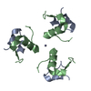
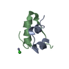
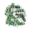
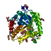
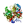
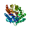
- PDB-1j73: Crystal structure of an unstable insulin analog with native activity. -
+ Open data

- Basic information
| Entry | Database: PDB / ID: 1j73 | ||||||
|---|---|---|---|---|---|---|---|
| Title | Crystal structure of an unstable insulin analog with native activity. | ||||||
 Components Components |
| ||||||
 Keywords Keywords | HORMONE/GROWTH FACTOR / A8-Diaminobutyric Acid human insulin / hormone-receptor interface / HORMONE-GROWTH FACTOR COMPLEX | ||||||
| Function / homology |  Function and homology information Function and homology informationnegative regulation of glycogen catabolic process / : / negative regulation of fatty acid metabolic process / Signaling by Insulin receptor / IRS activation / Insulin processing / regulation of protein secretion / positive regulation of peptide hormone secretion / negative regulation of feeding behavior / negative regulation of acute inflammatory response ...negative regulation of glycogen catabolic process / : / negative regulation of fatty acid metabolic process / Signaling by Insulin receptor / IRS activation / Insulin processing / regulation of protein secretion / positive regulation of peptide hormone secretion / negative regulation of feeding behavior / negative regulation of acute inflammatory response / Regulation of gene expression in beta cells / positive regulation of respiratory burst / alpha-beta T cell activation / Synthesis, secretion, and deacylation of Ghrelin / negative regulation of protein secretion / positive regulation of dendritic spine maintenance / negative regulation of gluconeogenesis / fatty acid homeostasis / positive regulation of glycogen biosynthetic process / positive regulation of insulin receptor signaling pathway / Signal attenuation / FOXO-mediated transcription of oxidative stress, metabolic and neuronal genes / negative regulation of lipid catabolic process / negative regulation of respiratory burst involved in inflammatory response / positive regulation of lipid biosynthetic process / negative regulation of oxidative stress-induced intrinsic apoptotic signaling pathway / nitric oxide-cGMP-mediated signaling / regulation of protein localization to plasma membrane / positive regulation of nitric-oxide synthase activity / transport vesicle / Insulin receptor recycling / COPI-mediated anterograde transport / negative regulation of reactive oxygen species biosynthetic process / positive regulation of brown fat cell differentiation / insulin-like growth factor receptor binding / NPAS4 regulates expression of target genes / neuron projection maintenance / positive regulation of mitotic nuclear division / endoplasmic reticulum-Golgi intermediate compartment membrane / Insulin receptor signalling cascade / positive regulation of glycolytic process / endosome lumen / acute-phase response / positive regulation of cytokine production / positive regulation of D-glucose import across plasma membrane / insulin receptor binding / positive regulation of long-term synaptic potentiation / positive regulation of protein secretion / positive regulation of cell differentiation / wound healing / Regulation of insulin secretion / positive regulation of neuron projection development / hormone activity / negative regulation of protein catabolic process / positive regulation of protein localization to nucleus / regulation of synaptic plasticity / glucose metabolic process / Golgi lumen / cognition / vasodilation / insulin receptor signaling pathway / glucose homeostasis / cell-cell signaling / regulation of protein localization / PI5P, PP2A and IER3 Regulate PI3K/AKT Signaling / positive regulation of cell growth / protease binding / secretory granule lumen / positive regulation of MAPK cascade / positive regulation of canonical NF-kappaB signal transduction / positive regulation of phosphatidylinositol 3-kinase/protein kinase B signal transduction / positive regulation of cell migration / endoplasmic reticulum lumen / G protein-coupled receptor signaling pathway / Amyloid fiber formation / receptor ligand activity / Golgi membrane / negative regulation of gene expression / positive regulation of cell population proliferation / positive regulation of gene expression / regulation of DNA-templated transcription / : / extracellular region / identical protein binding Similarity search - Function | ||||||
| Method |  X-RAY DIFFRACTION / Resolution: 2 Å X-RAY DIFFRACTION / Resolution: 2 Å | ||||||
 Authors Authors | Wan, Z. / Zhao, M. / Nakagawa, S. / Jia, W. / Weiss, M.A. | ||||||
 Citation Citation | Journal: J.Mol.Biol. / Year: 2002 Title: Non-standard insulin design: structure-activity relationships at the periphery of the insulin receptor. Authors: Weiss, M.A. / Wan, Z. / Zhao, M. / Chu, Y.C. / Nakagawa, S.H. / Burke, G.T. / Jia, W. / Hellmich, R. / Katsoyannis, P.G. #1: Journal: Proc.Natl.Acad.Sci.USA / Year: 2000Title: Diabetes-associated mutations in a beta-cell transcription factor destabilize an antiparallel "mini-zipper" in a dimerization interface Authors: Hua, Q.X. / Zhao, M. / Narayana, N. / Nakagawa, S.H. / Jia, W.H. / Weiss, M.A. #2: Journal: J.Mol.Biol. / Year: 1998Title: The Relationship Between Insulin Bioactivity and Structure in the NH2-terminal A-chain Helix Authors: Olsen, H.B. / Ludvigsen, S. / Kaarsholm, N.C. #3: Journal: J.Mol.Biol. / Year: 1996Title: Mapping the Functional Surface of Insulin by Design: Structure and Function of a Novel A-chain Analogue Authors: Hua, Q.X. / Hu, S.Q. / Frank, B.H. / Jia, W.H. / Chu, Y.C. / Wang, S.H. / Burke, G.T. / Kaysoyannis, P.G. / Weiss, M.A. #4: Journal: Biophys.Chem. / Year: 1994Title: A Proposed Interaction Model of the Insulin Molecule with its Receptor Authors: Liang, D.C. / Chang, W.R. / Wan, Z.L. / Vijayan, N.M. | ||||||
| History |
| ||||||
| Remark 400 | COMPOUND THE CONFORMATIONS OF TWO MONOMERS ARE DIFFERENT AS THE RESULT OF A CHANGE IN CONFORMATION ...COMPOUND THE CONFORMATIONS OF TWO MONOMERS ARE DIFFERENT AS THE RESULT OF A CHANGE IN CONFORMATION OF THE FIRST EIGHT RESIDUES OF THE B-CHAINS. |
- Structure visualization
| Structure viewer | Molecule: MolmilJmol/JSmol |
|---|
- Downloads & links
-Download
| PDBx/mmCIF format | 1j73.cif.gz | 34.8 KB | Display | PDBx/mmCIF format |
|---|---|---|---|---|
| PDB format | pdb1j73.ent.gz | 24 KB | Display | PDB format |
| PDBx/mmJSON format | 1j73.json.gz | Tree view | PDBx/mmJSON format | |
| Others |  Other downloads Other downloads |
-Validation report
| Arichive directory | https://data.pdbj.org/pub/pdb/validation_reports/j7/1j73ftp://data.pdbj.org/pub/pdb/validation_reports/j7/1j73 | HTTPS FTP |
|---|
-Related structure data


-Links
 PDBj
PDBj


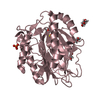
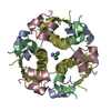
- Assembly
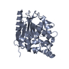
| Deposited unit | 
| |||||||||
|---|---|---|---|---|---|---|---|---|---|---|
| 1 | 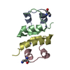
| |||||||||
| Unit cell |
| |||||||||
| Components on special symmetry positions |
|
-Components
| #1: Protein/peptide | Mass: 2382.713 Da / Num. of mol.: 2 / Mutation: T8(DAB) / Source method: obtained synthetically Details: The protein was chemically synthesized. The sequence is naturally found in Homo sapiens(human). References: UniProt: P01308 #2: Protein/peptide | Mass: 3433.953 Da / Num. of mol.: 2 / Source method: obtained synthetically Details: The protein was chemically synthesized. The sequence is naturally found in Homo sapiens(human). References: UniProt: P01308 #3: Chemical |   Mass: 65.409 Da / Num. of mol.: 2 / Source method: obtained synthetically / Formula: Zn Mass: 65.409 Da / Num. of mol.: 2 / Source method: obtained synthetically / Formula: Zn#4: Water | ChemComp-HOH / |  Mass: 18.015 Da / Num. of mol.: 95 / Source method: isolated from a natural source / Formula: H2O Mass: 18.015 Da / Num. of mol.: 95 / Source method: isolated from a natural source / Formula: H2OHas protein modification | Y | |
|---|
-Experimental details
-Experiment
| Experiment | Method: X-RAY DIFFRACTION / Number of used crystals: 1 |
|---|
- Sample preparation
| Crystal | Density Matthews: 1.91 Å3/Da / Density % sol: 35.75 % | ||||||||||||||||||||||||||||||||||||||||||||||||||||||||
|---|---|---|---|---|---|---|---|---|---|---|---|---|---|---|---|---|---|---|---|---|---|---|---|---|---|---|---|---|---|---|---|---|---|---|---|---|---|---|---|---|---|---|---|---|---|---|---|---|---|---|---|---|---|---|---|---|---|
| Crystal grow | Temperature: 298 K / Method: vapor diffusion, hanging drop / pH: 6.4 Details: Tris, phonel, acetone, sodium citrate, pH 6.4, VAPOR DIFFUSION, HANGING DROP, temperature 298K | ||||||||||||||||||||||||||||||||||||||||||||||||||||||||
| Crystal grow | *PLUS pH: 7.8 | ||||||||||||||||||||||||||||||||||||||||||||||||||||||||
| Components of the solutions | *PLUS
|
-Data collection
| Diffraction | Mean temperature: 100 K |
|---|---|
| Diffraction source | Source: ROTATING ANODE / Type: RIGAKU RU200 / Wavelength: 1.5418 Å |
| Detector | Type: RIGAKU RAXIS II / Detector: IMAGE PLATE / Date: Apr 26, 1998 / Details: mirrors |
| Radiation | Monochromator: Mirrors / Protocol: SINGLE WAVELENGTH / Monochromatic (M) / Laue (L): M / Scattering type: x-ray |
| Radiation wavelength | Wavelength: 1.5418 Å / Relative weight: 1 |
| Reflection | Resolution: 2→10 Å / Num. all: 5681 / Num. obs: 5448 / % possible obs: 95 % / Observed criterion σ(F): 2 / Observed criterion σ(I): 1 / Redundancy: 3 % / Rmerge(I) obs: 0.05 |
| Reflection shell | Resolution: 2→2.03 Å / Redundancy: 611 % / Rmerge(I) obs: 0.097 / % possible all: 100 |
| Reflection | *PLUS % possible obs: 93 % / Rmerge(I) obs: 0.056 |
- Processing
| Software |
| |||||||||||||||||||||||||
|---|---|---|---|---|---|---|---|---|---|---|---|---|---|---|---|---|---|---|---|---|---|---|---|---|---|---|
| Refinement | Resolution: 2→10 Å / σ(F): 2 / σ(I): 1
| |||||||||||||||||||||||||
| Refinement step | Cycle: LAST / Resolution: 2→10 Å
| |||||||||||||||||||||||||
| Software | *PLUS Name: X-PLOR / Version: 3.1 / Classification: refinement | |||||||||||||||||||||||||
| Refinement | *PLUS Highest resolution: 2 Å / Lowest resolution: 10 Å / σ(F): 2 / % reflection Rfree: 9.2 % / Rfactor obs: 0.205 | |||||||||||||||||||||||||
| Solvent computation | *PLUS | |||||||||||||||||||||||||
| Displacement parameters | *PLUS | |||||||||||||||||||||||||
| Refine LS restraints | *PLUS
|
