 Movie
Movie Controller

[English] 日本語
 Yorodumi
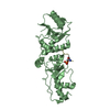
- PDB-2iwe: Structure of a cavity mutant (H117G) of Pseudomonas aeruginosa azurin -
+ Open data

- Basic information
| Entry | Database: PDB / ID: 2iwe | ||||||
|---|---|---|---|---|---|---|---|
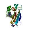
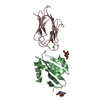
| Title | Structure of a cavity mutant (H117G) of Pseudomonas aeruginosa azurin | ||||||
 Components Components | AZURIN | ||||||
 Keywords Keywords | ELECTRON TRANSPORT / BLUE COPPER PROTEIN / REDOX PROTEIN / METAL-BINDING / AZURIN / TRANSPORT / PERIPLASMIC | ||||||
| Function / homology |  Function and homology information Function and homology informationtransition metal ion binding / periplasmic space / electron transfer activity / copper ion binding / zinc ion binding / identical protein binding / plasma membrane Similarity search - Function | ||||||
| Biological species |   PSEUDOMONAS AERUGINOSA (bacteria) PSEUDOMONAS AERUGINOSA (bacteria) | ||||||
| Method |  X-RAY DIFFRACTION / MOLECULAR REPLACEMENT / Resolution: 2.83 Å X-RAY DIFFRACTION / MOLECULAR REPLACEMENT / Resolution: 2.83 Å | ||||||
 Authors Authors | De Jongh, T.E. / Van Roon, A.M.M. / Prudencio, M. / Ubbink, M. / Canters, G.W. | ||||||
 Citation Citation | Journal: Eur.J.Inorg.Chem. / Year: 2006 Title: Click-Chemistry with an Active Site Variant of Azurin Authors: De Jongh, T.E. / Van Roon, A.M.M. / Prudencio, M. / Ubbink, M. / Canters, G.W. | ||||||
| History |
|
- Structure visualization
| Structure viewer | Molecule: MolmilJmol/JSmol |
|---|
- Downloads & links
-Download
| PDBx/mmCIF format | 2iwe.cif.gz | 109.9 KB | Display | PDBx/mmCIF format |
|---|---|---|---|---|
| PDB format | pdb2iwe.ent.gz | 86 KB | Display | PDB format |
| PDBx/mmJSON format | 2iwe.json.gz | Tree view | PDBx/mmJSON format | |
| Others |  Other downloads Other downloads |
-Validation report
| Summary document | 2iwe_validation.pdf.gz | 464 KB | Display | wwPDB validaton report |
|---|---|---|---|---|
| Full document | 2iwe_full_validation.pdf.gz | 473.9 KB | Display | |
| Data in XML | 2iwe_validation.xml.gz | 20.6 KB | Display | |
| Data in CIF | 2iwe_validation.cif.gz | 26.9 KB | Display | |
| Arichive directory | https://data.pdbj.org/pub/pdb/validation_reports/iw/2iweftp://data.pdbj.org/pub/pdb/validation_reports/iw/2iwe | HTTPS FTP |
-Related structure data
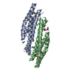
| Related structure data | 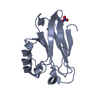 1e67S S: Starting model for refinement |
|---|---|
| Similar structure data |


-Links
 PDBj
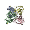
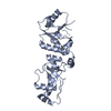
PDBj- Assembly
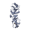
| Deposited unit | 
| ||||||||||||||||||||||||||||||||||||||||||||||
|---|---|---|---|---|---|---|---|---|---|---|---|---|---|---|---|---|---|---|---|---|---|---|---|---|---|---|---|---|---|---|---|---|---|---|---|---|---|---|---|---|---|---|---|---|---|---|---|
| 1 | 
| ||||||||||||||||||||||||||||||||||||||||||||||
| 2 | 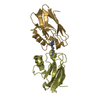
| ||||||||||||||||||||||||||||||||||||||||||||||
| Unit cell |
| ||||||||||||||||||||||||||||||||||||||||||||||
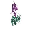
| Noncrystallographic symmetry (NCS) | NCS domain:
NCS domain segments: Component-ID: 1 / Ens-ID: 1 / Beg auth comp-ID: ALA / Beg label comp-ID: ALA / End auth comp-ID: PHE / End label comp-ID: PHE / Refine code: 1 / Auth seq-ID: 1 - 114 / Label seq-ID: 1 - 114
NCS oper:
|
-Components
| #1: Protein | Mass: 13880.704 Da / Num. of mol.: 4 / Mutation: YES Source method: isolated from a genetically manipulated source Details: DIMERIZED BY COORDINATION OF A BIFUNCTIONAL LIGAND WIRE 1,6-DI(IMIDAZOL-1-YL)HEXANE Source: (gene. exp.) PSEUDOMONAS AERUGINOSA (bacteria) / Plasmid: PGK22 (PUC18 DERIVED) / Production host: #2: Chemical | ChemComp-ZN /   Mass: 65.409 Da / Num. of mol.: 4 / Source method: obtained synthetically / Formula: Zn Mass: 65.409 Da / Num. of mol.: 4 / Source method: obtained synthetically / Formula: Zn#3: Chemical |   Mass: 218.298 Da / Num. of mol.: 2 / Source method: obtained synthetically / Formula: C12H18N4 Mass: 218.298 Da / Num. of mol.: 2 / Source method: obtained synthetically / Formula: C12H18N4#4: Water | ChemComp-HOH / |  Mass: 18.015 Da / Num. of mol.: 3 / Source method: isolated from a natural source / Formula: H2O Mass: 18.015 Da / Num. of mol.: 3 / Source method: isolated from a natural source / Formula: H2OCompound details | AZURIN, FOUND IN BACTERIA, IS THOUGHT TO TRANSFER ELECTRONS FROM CYTOCHROME C551 TO CYTOCHROME ...AZURIN, FOUND IN BACTERIA, IS THOUGHT TO TRANSFER ELECTRONS FROM CYTOCHROME | Has protein modification | Y | |
|---|
-Experimental details
-Experiment
| Experiment | Method: X-RAY DIFFRACTION / Number of used crystals: 1 |
|---|
- Sample preparation
| Crystal | Density Matthews: 2.3 Å3/Da / Density % sol: 46 % |
|---|---|
| Crystal grow | pH: 8.5 Details: 100 MM TRIS-HCL PH 8.5 AND 20% (W/V) POLYETHYLENE GLYCOL (PEG) 8000 |
-Data collection
| Diffraction | Mean temperature: 100 K |
|---|---|
| Diffraction source | Source: ROTATING ANODE / Type: ENRAF-NONIUS FR78 / Wavelength: 1.5418 |
| Detector | Type: MAR scanner 345 mm plate / Detector: IMAGE PLATE / Date: Mar 9, 2004 / Details: MIRRORS |
| Radiation | Monochromator: NI FILTER / Protocol: SINGLE WAVELENGTH / Monochromatic (M) / Laue (L): M / Scattering type: x-ray |
| Radiation wavelength | Wavelength: 1.5418 Å / Relative weight: 1 |
| Reflection | Resolution: 2.85→60 Å / Num. obs: 10566 / % possible obs: 88.2 % / Observed criterion σ(I): 1.8 / Redundancy: 2.7 % / Biso Wilson estimate: 62.5 Å2 / Rmerge(I) obs: 0.09 / Net I/σ(I): 9.4 |
| Reflection shell | Resolution: 2.85→2.95 Å / Redundancy: 1.6 % / Rmerge(I) obs: 0.34 / Mean I/σ(I) obs: 1.8 / % possible all: 36 |
- Processing
| Software |
| ||||||||||||||||||||||||||||||||||||||||||||||||||||||||||||||||||||||||||||||||||||||||||||||||||||||||||||||||||||||||||||||||||||||||||||||||||||||||||||||||||||||||||||||||||||||
|---|---|---|---|---|---|---|---|---|---|---|---|---|---|---|---|---|---|---|---|---|---|---|---|---|---|---|---|---|---|---|---|---|---|---|---|---|---|---|---|---|---|---|---|---|---|---|---|---|---|---|---|---|---|---|---|---|---|---|---|---|---|---|---|---|---|---|---|---|---|---|---|---|---|---|---|---|---|---|---|---|---|---|---|---|---|---|---|---|---|---|---|---|---|---|---|---|---|---|---|---|---|---|---|---|---|---|---|---|---|---|---|---|---|---|---|---|---|---|---|---|---|---|---|---|---|---|---|---|---|---|---|---|---|---|---|---|---|---|---|---|---|---|---|---|---|---|---|---|---|---|---|---|---|---|---|---|---|---|---|---|---|---|---|---|---|---|---|---|---|---|---|---|---|---|---|---|---|---|---|---|---|---|---|
| Refinement | Method to determine structure: MOLECULAR REPLACEMENT Starting model: PDB ENTRY 1E67 Resolution: 2.83→61.66 Å / Cor.coef. Fo:Fc: 0.937 / Cor.coef. Fo:Fc free: 0.908 / SU B: 44.56 / SU ML: 0.378 / TLS residual ADP flag: LIKELY RESIDUAL / Cross valid method: THROUGHOUT / σ(F): 2 / ESU R Free: 0.436 / Stereochemistry target values: MAXIMUM LIKELIHOOD Details: HYDROGENS HAVE BEEN ADDED IN THE RIDING POSITIONS. THE LOOP COMPRISING RESIDUES 116-121 IN CHAIN A IS MODELED IN A DOUBLE CONFORMATION.
| ||||||||||||||||||||||||||||||||||||||||||||||||||||||||||||||||||||||||||||||||||||||||||||||||||||||||||||||||||||||||||||||||||||||||||||||||||||||||||||||||||||||||||||||||||||||
| Solvent computation | Ion probe radii: 0.8 Å / VDW probe radii: 1.2 Å / Solvent model: BABINET MODEL PLUS MASK | ||||||||||||||||||||||||||||||||||||||||||||||||||||||||||||||||||||||||||||||||||||||||||||||||||||||||||||||||||||||||||||||||||||||||||||||||||||||||||||||||||||||||||||||||||||||
| Displacement parameters | Biso mean: 30.6 Å2
| ||||||||||||||||||||||||||||||||||||||||||||||||||||||||||||||||||||||||||||||||||||||||||||||||||||||||||||||||||||||||||||||||||||||||||||||||||||||||||||||||||||||||||||||||||||||
| Refinement step | Cycle: LAST / Resolution: 2.83→61.66 Å
| ||||||||||||||||||||||||||||||||||||||||||||||||||||||||||||||||||||||||||||||||||||||||||||||||||||||||||||||||||||||||||||||||||||||||||||||||||||||||||||||||||||||||||||||||||||||
| Refine LS restraints |
|