 Movie
Movie Controller


+ Open data

- Basic information
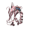
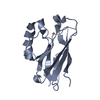
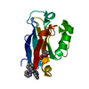
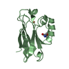
| Entry | Database: PDB / ID: 1i53 | |||||||||
|---|---|---|---|---|---|---|---|---|---|---|
| Title | RE(I)-TRICARBONYL DIIMINE (Q107H)) AZURIN | |||||||||
 Components Components | AZURIN | |||||||||
 Keywords Keywords | ELECTRON TRANSPORT / azurin / rhenium / electron transfer / tyrosyl and tryptophan radical | |||||||||
| Function / homology |  Function and homology information Function and homology informationtransition metal ion binding / electron transfer activity / periplasmic space / copper ion binding / zinc ion binding / identical protein binding / plasma membrane Similarity search - Function | |||||||||
| Biological species |   Pseudomonas aeruginosa (bacteria) Pseudomonas aeruginosa (bacteria) | |||||||||
| Method |  X-RAY DIFFRACTION / MOLECULAR REPLACEMENT / Resolution: 1.8 Å X-RAY DIFFRACTION / MOLECULAR REPLACEMENT / Resolution: 1.8 Å | |||||||||
 Authors Authors | Di Bilio, A.J. / Crane, B.R. / Wehbi, W.A. / Kiser, C.N. / Abu-Omar, M.M. / Carlos, R.M. / Richards, J.H. / Winkler, J.R. / Gray, H.B. | |||||||||
 Citation Citation | Journal: J.Am.Chem.Soc. / Year: 2001 Title: Properties of photogenerated tryptophan and tyrosyl radicals in structurally characterized proteins containing rhenium(I) tricarbonyl diimines. Authors: Di Bilio, A.J. / Crane, B.R. / Wehbi, W.A. / Kiser, C.N. / Abu-Omar, M.M. / Carlos, R.M. / Richards, J.H. / Winkler, J.R. / Gray, H.B. | |||||||||
| History |
|
- Structure visualization
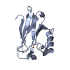
| Structure viewer | Molecule: MolmilJmol/JSmol |
|---|
- Downloads & links
-Download
| PDBx/mmCIF format | 1i53.cif.gz | 68.1 KB | Display | PDBx/mmCIF format |
|---|---|---|---|---|
| PDB format | pdb1i53.ent.gz | 49.8 KB | Display | PDB format |
| PDBx/mmJSON format | 1i53.json.gz | Tree view | PDBx/mmJSON format | |
| Others |  Other downloads Other downloads |
-Validation report
| Arichive directory | https://data.pdbj.org/pub/pdb/validation_reports/i5/1i53ftp://data.pdbj.org/pub/pdb/validation_reports/i5/1i53 | HTTPS FTP |
|---|
-Related structure data
| Related structure data |  1bexS S: Starting model for refinement |
|---|---|
| Similar structure data |


-Links
 PDBj
PDBj- Assembly
| Deposited unit | 
| ||||||||
|---|---|---|---|---|---|---|---|---|---|
| 1 | 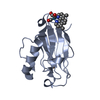
| ||||||||
| 2 | 
| ||||||||
| Unit cell |
|
-Components
| #1: Protein | Mass: 13960.814 Da / Num. of mol.: 2 / Mutation: Q107H Source method: isolated from a genetically manipulated source Source: (gene. exp.) Pseudomonas aeruginosa (bacteria) / Plasmid: PET9A/ASA / Species (production host): Escherichia coli / Production host: #2: Chemical |   Mass: 63.546 Da / Num. of mol.: 2 / Source method: obtained synthetically / Formula: Cu Mass: 63.546 Da / Num. of mol.: 2 / Source method: obtained synthetically / Formula: Cu#3: Chemical |   Mass: 270.237 Da / Num. of mol.: 2 / Source method: obtained synthetically / Formula: C3O3Re Mass: 270.237 Da / Num. of mol.: 2 / Source method: obtained synthetically / Formula: C3O3Re#4: Chemical |   Mass: 208.258 Da / Num. of mol.: 2 / Source method: obtained synthetically / Formula: C14H12N2 Mass: 208.258 Da / Num. of mol.: 2 / Source method: obtained synthetically / Formula: C14H12N2#5: Water | ChemComp-HOH / |  Mass: 18.015 Da / Num. of mol.: 239 / Source method: isolated from a natural source / Formula: H2O Mass: 18.015 Da / Num. of mol.: 239 / Source method: isolated from a natural source / Formula: H2OHas protein modification | Y | |
|---|
-Experimental details
-Experiment
| Experiment | Method: X-RAY DIFFRACTION / Number of used crystals: 1 |
|---|
- Sample preparation
| Crystal | Density Matthews: 2.33 Å3/Da / Density % sol: 47.29 % | |||||||||||||||||||||||||
|---|---|---|---|---|---|---|---|---|---|---|---|---|---|---|---|---|---|---|---|---|---|---|---|---|---|---|
| Crystal grow | Temperature: 295 K / Method: vapor diffusion, hanging drop / pH: 8 Details: CuCl2, Imidazole, LiNo3, PEG 8K, pH 8.0, VAPOR DIFFUSION, HANGING DROP, temperature 295K | |||||||||||||||||||||||||
| Crystal grow | *PLUS pH: 7.5 / Method: vapor diffusion | |||||||||||||||||||||||||
| Components of the solutions | *PLUS
|
-Data collection
| Diffraction | Mean temperature: 100 K |
|---|---|
| Diffraction source | Source: ROTATING ANODE / Type: RIGAKU RU300 / Wavelength: 1.5418 Å |
| Detector | Type: RIGAKU RAXIS IV / Detector: IMAGE PLATE / Date: Jun 29, 2000 / Details: mirrors |
| Radiation | Protocol: SINGLE WAVELENGTH / Monochromatic (M) / Laue (L): M / Scattering type: x-ray |
| Radiation wavelength | Wavelength: 1.5418 Å / Relative weight: 1 |
| Reflection | Resolution: 1.8→30 Å / Num. all: 20201 / Num. obs: 45216 / % possible obs: 86 % / Observed criterion σ(F): 1 / Observed criterion σ(I): 1 / Redundancy: 2.2 % / Biso Wilson estimate: 19.9 Å2 / Rmerge(I) obs: 0.061 / Net I/σ(I): 20.4 |
| Reflection shell | Resolution: 1.8→1.89 Å / Redundancy: 2.2 % / Rmerge(I) obs: 0.112 / % possible all: 83 |
| Reflection | *PLUS Num. all: 45216 / Num. obs: 20201 |
- Processing
| Software |
| ||||||||||||||||||||||||||||||||||||||||
|---|---|---|---|---|---|---|---|---|---|---|---|---|---|---|---|---|---|---|---|---|---|---|---|---|---|---|---|---|---|---|---|---|---|---|---|---|---|---|---|---|---|
| Refinement | Method to determine structure: MOLECULAR REPLACEMENT Starting model: 1BEX Resolution: 1.8→24.56 Å / Rfactor Rfree error: 0.006 / Data cutoff high absF: 1144283.98 / Data cutoff low absF: 0 / Isotropic thermal model: RESTRAINED / σ(F): 0 / σ(I): 0 Stereochemistry target values: Engh and Huber + CSB model complex values for Re label
| ||||||||||||||||||||||||||||||||||||||||
| Solvent computation | Solvent model: FLAT MODEL / Bsol: 42.77 Å2 / ksol: 0.362 e/Å3 | ||||||||||||||||||||||||||||||||||||||||
| Displacement parameters | Biso mean: 20.9 Å2
| ||||||||||||||||||||||||||||||||||||||||
| Refine analyze |
| ||||||||||||||||||||||||||||||||||||||||
| Refinement step | Cycle: LAST / Resolution: 1.8→24.56 Å
| ||||||||||||||||||||||||||||||||||||||||
| Refine LS restraints |
| ||||||||||||||||||||||||||||||||||||||||
| LS refinement shell | Resolution: 1.8→1.86 Å / Rfactor Rfree error: 0.027 / Total num. of bins used: 10
| ||||||||||||||||||||||||||||||||||||||||
| Xplor file |
| ||||||||||||||||||||||||||||||||||||||||
| Software | *PLUS Name: CNS / Version: 1 / Classification: refinement | ||||||||||||||||||||||||||||||||||||||||
| Refinement | *PLUS σ(F): 0 / % reflection Rfree: 8 % | ||||||||||||||||||||||||||||||||||||||||
| Solvent computation | *PLUS | ||||||||||||||||||||||||||||||||||||||||
| Displacement parameters | *PLUS Biso mean: 20.9 Å2 | ||||||||||||||||||||||||||||||||||||||||
| Refine LS restraints | *PLUS
| ||||||||||||||||||||||||||||||||||||||||
| LS refinement shell | *PLUS Rfactor Rfree: 0.319 / % reflection Rfree: 7.4 % / Rfactor Rwork: 0.311 |
