 Movie
Movie Controller

[English] 日本語
 Yorodumi
- PDB-3in0: Crystal structure of the F114P/M121Q variant of Pseudomonas aerug... -
+ Open data

- Basic information
| Entry | Database: PDB / ID: 3in0 | ||||||
|---|---|---|---|---|---|---|---|
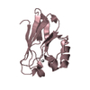
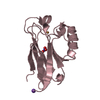
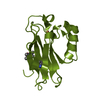
| Title | Crystal structure of the F114P/M121Q variant of Pseudomonas aeruginosa azurin in the Cu(II) state | ||||||
 Components Components | Azurin | ||||||
 Keywords Keywords | ELECTRON TRANSPORT / CUPREDOXIN / AZURIN / GREEK KEY / BETA BARREL / ELECTRON TRANSFER / Copper / Disulfide bond / Metal-binding / Periplasm / Transport | ||||||
| Function / homology |  Function and homology information Function and homology informationtransition metal ion binding / electron transfer activity / periplasmic space / copper ion binding / zinc ion binding / identical protein binding / plasma membrane Similarity search - Function | ||||||
| Biological species |   Pseudomonas aeruginosa (bacteria) Pseudomonas aeruginosa (bacteria) | ||||||
| Method |  X-RAY DIFFRACTION / SYNCHROTRON / AB INITIO / Resolution: 2.35 Å X-RAY DIFFRACTION / SYNCHROTRON / AB INITIO / Resolution: 2.35 Å | ||||||
 Authors Authors | Gao, Y.G. / Robinson, H. | ||||||
 Citation Citation | Journal: Nature / Year: 2009 Title: Rationally tuning the reduction potential of a single cupredoxin beyond the natural range. Authors: Marshall, N.M. / Garner, D.K. / Wilson, T.D. / Gao, Y.G. / Robinson, H. / Nilges, M.J. / Lu, Y. | ||||||
| History |
|
- Structure visualization
| Structure viewer | Molecule: MolmilJmol/JSmol |
|---|
- Downloads & links
-Download
| PDBx/mmCIF format | 3in0.cif.gz | 108.8 KB | Display | PDBx/mmCIF format |
|---|---|---|---|---|
| PDB format | pdb3in0.ent.gz | 84.4 KB | Display | PDB format |
| PDBx/mmJSON format | 3in0.json.gz | Tree view | PDBx/mmJSON format | |
| Others |  Other downloads Other downloads |
-Validation report
| Arichive directory | https://data.pdbj.org/pub/pdb/validation_reports/in/3in0ftp://data.pdbj.org/pub/pdb/validation_reports/in/3in0 | HTTPS FTP |
|---|
-Related structure data


-Links
 PDBj
PDBj- Assembly
| Deposited unit | 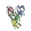
| ||||||||
|---|---|---|---|---|---|---|---|---|---|
| 1 | 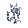
| ||||||||
| 2 | 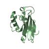
| ||||||||
| 3 | 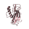
| ||||||||
| 4 | 
| ||||||||
| Unit cell |
|
-Components
| #1: Protein | Mass: 13908.672 Da / Num. of mol.: 4 / Mutation: F114P, M121Q Source method: isolated from a genetically manipulated source Details: Oxidized F114P/M121Q Azurin / Source: (gene. exp.) Pseudomonas aeruginosa (bacteria) / Gene: azu, PA4922 / Plasmid: pET9a / Production host: #2: Chemical | ChemComp-CU /   Mass: 63.546 Da / Num. of mol.: 4 / Source method: obtained synthetically / Formula: Cu Mass: 63.546 Da / Num. of mol.: 4 / Source method: obtained synthetically / Formula: Cu#3: Water | ChemComp-HOH / |  Mass: 18.015 Da / Num. of mol.: 134 / Source method: isolated from a natural source / Formula: H2O Mass: 18.015 Da / Num. of mol.: 134 / Source method: isolated from a natural source / Formula: H2OHas protein modification | Y | |
|---|
-Experimental details
-Experiment
| Experiment | Method: X-RAY DIFFRACTION / Number of used crystals: 1 |
|---|
- Sample preparation
| Crystal | Density Matthews: 2.18 Å3/Da / Density % sol: 43.51 % |
|---|---|
| Crystal grow | Temperature: 277 K / Method: vapor diffusion, hanging drop / pH: 6 Details: PEG 8000, 0.08M NaOAc, 0.2M Lithium nitrate, 0.2M Calcium chloride, pH 6.0, VAPOR DIFFUSION, HANGING DROP, temperature 277K |
-Data collection
| Diffraction | Mean temperature: 100 K |
|---|---|
| Diffraction source | Source: SYNCHROTRON / Site: NSLS  / Beamline: X29A / Wavelength: 1.1 Å / Beamline: X29A / Wavelength: 1.1 Å |
| Detector | Type: ADSC QUANTUM 315 / Detector: CCD Details: Cryogenically cooled double crystal monochromator with horizontally focusing sagitally bent second mono crystal |
| Radiation | Monochromator: Si(111) / Protocol: SINGLE WAVELENGTH / Monochromatic (M) / Laue (L): M / Scattering type: x-ray |
| Radiation wavelength | Wavelength: 1.1 Å / Relative weight: 1 |
| Reflection | Resolution: 2→50 Å / Num. obs: 29479 / Redundancy: 3.8 % / Rmerge(I) obs: 0.065 |
| Reflection shell | Resolution: 2→2.1 Å |
- Processing
| Software |
| |||||||||||||||||||||||||||||||||
|---|---|---|---|---|---|---|---|---|---|---|---|---|---|---|---|---|---|---|---|---|---|---|---|---|---|---|---|---|---|---|---|---|---|---|
| Refinement | Method to determine structure: AB INITIO / Resolution: 2.35→10 Å / Num. parameters: 12215 / Num. restraintsaints: 16036 / Cross valid method: FREE R / Stereochemistry target values: ENGH AND HUBER Details: ANISOTROPIC SCALING APPLIED BY THE METHOD OF PARKIN, MOEZZI & HOPE, J.APPL.CRYST.28(1995)53-56
| |||||||||||||||||||||||||||||||||
| Refine analyze | Num. disordered residues: 0 / Occupancy sum hydrogen: 0 / Occupancy sum non hydrogen: 4022 | |||||||||||||||||||||||||||||||||
| Refinement step | Cycle: LAST / Resolution: 2.35→10 Å
| |||||||||||||||||||||||||||||||||
| Refine LS restraints |
|
