 Movie
Movie Controller

[English] 日本語
 Yorodumi
- PDB-1gr7: Crystal structure of the double mutant Cys3Ser/Ser100Pro from Pse... -
+ Open data

- Basic information
| Entry | Database: PDB / ID: 1gr7 | ||||||
|---|---|---|---|---|---|---|---|
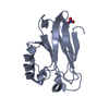
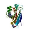
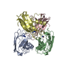
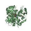
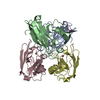
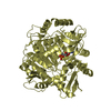
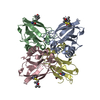
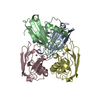
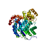
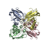
| Title | Crystal structure of the double mutant Cys3Ser/Ser100Pro from Pseudomonas Aeruginosa at 1.8 A resolution | ||||||
 Components Components | AZURIN | ||||||
 Keywords Keywords | ELECTRON TRANSPORT | ||||||
| Function / homology |  Function and homology information Function and homology informationtransition metal ion binding / electron transfer activity / periplasmic space / copper ion binding / zinc ion binding / identical protein binding / plasma membrane Similarity search - Function | ||||||
| Biological species |   PSEUDOMONAS AERUGINOSA (bacteria) PSEUDOMONAS AERUGINOSA (bacteria) | ||||||
| Method |  X-RAY DIFFRACTION / MOLECULAR REPLACEMENT / Resolution: 1.8 Å X-RAY DIFFRACTION / MOLECULAR REPLACEMENT / Resolution: 1.8 Å | ||||||
 Authors Authors | Okvist, M. / Bonander, N. / Sandberg, A. / Karlsson, B.G. / Krengel, U. / Xue, Y. / Sjolin, L. | ||||||
 Citation Citation | Journal: Biochim.Biophys.Acta / Year: 2002 Title: Crystal structure of the double azurin mutant Cys3Ser/Ser100Pro from Pseudomonas aeruginosa at 1.8 A resolution: its folding-unfolding energy and unfolding kinetics. Authors: Okvist, M. / Bonander, N. / Sandberg, A. / Karlsson, B.G. / Krengel, U. / Xue, Y. / Sjolin, L. | ||||||
| History |
|
- Structure visualization
| Structure viewer | Molecule: MolmilJmol/JSmol |
|---|
- Downloads & links
-Download
| PDBx/mmCIF format | 1gr7.cif.gz | 110.2 KB | Display | PDBx/mmCIF format |
|---|---|---|---|---|
| PDB format | pdb1gr7.ent.gz | 86.1 KB | Display | PDB format |
| PDBx/mmJSON format | 1gr7.json.gz | Tree view | PDBx/mmJSON format | |
| Others |  Other downloads Other downloads |
-Validation report
| Arichive directory | https://data.pdbj.org/pub/pdb/validation_reports/gr/1gr7ftp://data.pdbj.org/pub/pdb/validation_reports/gr/1gr7 | HTTPS FTP |
|---|
-Related structure data
| Related structure data | |
|---|---|
| Similar structure data |


-Links
 PDBj
PDBj- Assembly
| Deposited unit | 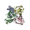
| ||||||||||||||||
|---|---|---|---|---|---|---|---|---|---|---|---|---|---|---|---|---|---|
| 1 |
| ||||||||||||||||
| Unit cell |
| ||||||||||||||||
| Noncrystallographic symmetry (NCS) | NCS oper:
| ||||||||||||||||
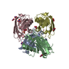
| Details | THE MOLECULE FUNCTIONS AS A MONOMER |
-Components
| #1: Protein | Mass: 13971.771 Da / Num. of mol.: 4 / Mutation: YES Source method: isolated from a genetically manipulated source Details: OXIDIZED CYSTEINE AT POSITION 26. / Source: (gene. exp.) PSEUDOMONAS AERUGINOSA (bacteria) / Production host: #2: Chemical | ChemComp-CU /   Mass: 63.546 Da / Num. of mol.: 4 / Source method: obtained synthetically / Formula: Cu Mass: 63.546 Da / Num. of mol.: 4 / Source method: obtained synthetically / Formula: Cu#3: Water | ChemComp-HOH / |  Mass: 18.015 Da / Num. of mol.: 175 / Source method: isolated from a natural source / Formula: H2O Mass: 18.015 Da / Num. of mol.: 175 / Source method: isolated from a natural source / Formula: H2OHas protein modification | Y | Sequence details | ENGINEERED | |
|---|
-Experimental details
-Experiment
| Experiment | Method: X-RAY DIFFRACTION / Number of used crystals: 1 |
|---|
- Sample preparation
| Crystal | Density Matthews: 2.3 Å3/Da / Density % sol: 45 % | ||||||||||||||||||||||||||||
|---|---|---|---|---|---|---|---|---|---|---|---|---|---|---|---|---|---|---|---|---|---|---|---|---|---|---|---|---|---|
| Crystal grow | pH: 5.5 Details: 3.2 M AMMONIUM SULPHATE, 1.0 M LITHIUM NITRATE, 0.2 M ACETATE BUFFER PH 5.5 | ||||||||||||||||||||||||||||
| Crystal grow | *PLUS Method: vapor diffusion, hanging drop | ||||||||||||||||||||||||||||
| Components of the solutions | *PLUS
|
-Data collection
| Diffraction | Mean temperature: 293 K |
|---|---|
| Diffraction source | Source: ROTATING ANODE / Type: RIGAKU RU200 / Wavelength: 1.5418 |
| Detector | Type: MSC / Detector: IMAGE PLATE |
| Radiation | Monochromator: GRAPHITE CRYSTAL / Protocol: SINGLE WAVELENGTH / Monochromatic (M) / Laue (L): M / Scattering type: x-ray |
| Radiation wavelength | Wavelength: 1.5418 Å / Relative weight: 1 |
| Reflection | Resolution: 1.8→30 Å / Num. obs: 248852 / % possible obs: 89.2 % / Redundancy: 5.6 % / Rmerge(I) obs: 0.066 / Net I/σ(I): 10.7 |
| Reflection shell | Resolution: 1.8→1.86 Å / Redundancy: 2.9 % / Rmerge(I) obs: 0.46 / Mean I/σ(I) obs: 2.2 / % possible all: 53.7 |
| Reflection | *PLUS Num. obs: 42969 / Num. measured all: 248852 |
| Reflection shell | *PLUS Highest resolution: 1.8 Å / % possible obs: 53.7 % / Mean I/σ(I) obs: 2.1 |
- Processing
| Software |
| ||||||||||||||||||||
|---|---|---|---|---|---|---|---|---|---|---|---|---|---|---|---|---|---|---|---|---|---|
| Refinement | Method to determine structure: MOLECULAR REPLACEMENT / Resolution: 1.8→72.55 Å / SU B: 5.68947 / SU ML: 0.16785 / Cross valid method: THROUGHOUT / σ(F): 0 / ESU R Free: 0.12985 / Details: HYDROGENS HAVE BEEN ADDED IN THE RIDING POSITIONS
| ||||||||||||||||||||
| Refinement step | Cycle: LAST / Resolution: 1.8→72.55 Å
| ||||||||||||||||||||
| Refinement | *PLUS Lowest resolution: 72.5 Å / % reflection Rfree: 5 % / Rfactor obs: 0.175 / Rfactor Rfree: 0.208 / Rfactor Rwork: 0.175 | ||||||||||||||||||||
| Solvent computation | *PLUS | ||||||||||||||||||||
| Displacement parameters | *PLUS | ||||||||||||||||||||
| Refine LS restraints | *PLUS
|
