 Movie
Movie Controller


+ Open data

- Basic information
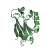
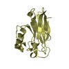
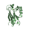
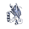
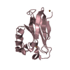
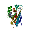
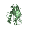
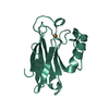
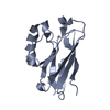
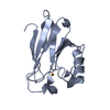
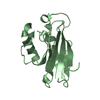
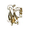
| Entry | Database: PDB / ID: 1etj | ||||||
|---|---|---|---|---|---|---|---|
| Title | AZURIN MUTANT WITH MET 121 REPLACED BY GLU | ||||||
 Components Components | AZURIN | ||||||
 Keywords Keywords | ELECTRON TRANSPORT / COPPER / PERIPLASMIC | ||||||
| Function / homology |  Function and homology information Function and homology informationtransition metal ion binding / electron transfer activity / periplasmic space / copper ion binding / zinc ion binding / identical protein binding / plasma membrane Similarity search - Function | ||||||
| Biological species |   Pseudomonas aeruginosa (bacteria) Pseudomonas aeruginosa (bacteria) | ||||||
| Method |  X-RAY DIFFRACTION / MOLECULAR REPLACEMENT / Resolution: 2.3 Å X-RAY DIFFRACTION / MOLECULAR REPLACEMENT / Resolution: 2.3 Å | ||||||
 Authors Authors | Karlsson, B.G. / Tsai, L.-C. / Nar, H. / Sanders-Loehr, J. / Bonander, N. / Langer, V. / Sjolin, L. | ||||||
 Citation Citation | Journal: Biochemistry / Year: 1997 Title: X-ray structure determination and characterization of the Pseudomonas aeruginosa azurin mutant Met121Glu. Authors: Karlsson, B.G. / Tsai, L.C. / Nar, H. / Sanders-Loehr, J. / Bonander, N. / Langer, V. / Sjolin, L. | ||||||
| History |
|
- Structure visualization
| Structure viewer | Molecule: MolmilJmol/JSmol |
|---|
- Downloads & links
-Download
| PDBx/mmCIF format | 1etj.cif.gz | 114 KB | Display | PDBx/mmCIF format |
|---|---|---|---|---|
| PDB format | pdb1etj.ent.gz | 88.4 KB | Display | PDB format |
| PDBx/mmJSON format | 1etj.json.gz | Tree view | PDBx/mmJSON format | |
| Others |  Other downloads Other downloads |
-Validation report
| Arichive directory | https://data.pdbj.org/pub/pdb/validation_reports/et/1etjftp://data.pdbj.org/pub/pdb/validation_reports/et/1etj | HTTPS FTP |
|---|
-Related structure data
| Similar structure data |
|---|


-Links
 PDBj
PDBj- Assembly
| Deposited unit | 
| ||||||||
|---|---|---|---|---|---|---|---|---|---|
| 1 | 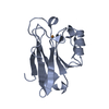
| ||||||||
| 2 | 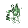
| ||||||||
| 3 | 
| ||||||||
| 4 | 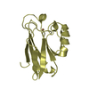
| ||||||||
| Unit cell |
|
-Components
| #1: Protein | Mass: 13959.717 Da / Num. of mol.: 4 / Mutation: M121E Source method: isolated from a genetically manipulated source Source: (gene. exp.) Pseudomonas aeruginosa (bacteria) / References: UniProt: P00282#2: Chemical | ChemComp-CU /   Mass: 63.546 Da / Num. of mol.: 4 / Source method: obtained synthetically / Formula: Cu Mass: 63.546 Da / Num. of mol.: 4 / Source method: obtained synthetically / Formula: Cu#3: Water | ChemComp-HOH / |  Mass: 18.015 Da / Num. of mol.: 308 / Source method: isolated from a natural source / Formula: H2O Mass: 18.015 Da / Num. of mol.: 308 / Source method: isolated from a natural source / Formula: H2OHas protein modification | Y | |
|---|
-Experimental details
-Experiment
| Experiment | Method: X-RAY DIFFRACTION / Number of used crystals: 1 |
|---|
- Sample preparation
| Crystal | Density Matthews: 2.25 Å3/Da / Density % sol: 55 % | ||||||||||||||||||||||||||||||
|---|---|---|---|---|---|---|---|---|---|---|---|---|---|---|---|---|---|---|---|---|---|---|---|---|---|---|---|---|---|---|---|
| Crystal grow | Method: vapor diffusion, hanging drop / pH: 6 Details: THE BLUISH WELL-FORMED PRISMATIC CRYSTALS OF THE TITLE PROTEIN WERE OBTAINED BY THE VAPOR-DIFFUSION HANGING-DROP TECHNIQUE FROM A SOLUTION CONTAINING 25% PEG4000, 0.24M CALCIUM DICHLORIDE ...Details: THE BLUISH WELL-FORMED PRISMATIC CRYSTALS OF THE TITLE PROTEIN WERE OBTAINED BY THE VAPOR-DIFFUSION HANGING-DROP TECHNIQUE FROM A SOLUTION CONTAINING 25% PEG4000, 0.24M CALCIUM DICHLORIDE AND 0.26M LITHIUM NITRATE BUFFER AT PH 6.0 AND AT THE TEMPERATURE OF 24 - 25 CENTIGRADE IN AROUND 10 DAYS., vapor diffusion - hanging drop | ||||||||||||||||||||||||||||||
| Crystal grow | *PLUS Method: unknown | ||||||||||||||||||||||||||||||
| Components of the solutions | *PLUS
|
-Data collection
| Diffraction | Mean temperature: 277 K |
|---|---|
| Diffraction source | Source: ROTATING ANODE / Type: RIGAKU RUH2R / Wavelength: 1.5418 |
| Detector | Type: ENRAF-NONIUS FAST / Detector: DIFFRACTOMETER |
| Radiation | Monochromator: GRAPHITE(002) / Monochromatic (M) / Laue (L): M / Scattering type: x-ray |
| Radiation wavelength | Wavelength: 1.5418 Å / Relative weight: 1 |
| Reflection | Resolution: 2.3→8 Å / Num. obs: 17964 / % possible obs: 75.7 % / Observed criterion σ(I): 2 / Redundancy: 2.77 % / Rmerge(I) obs: 0.115 / Rsym value: 0.115 |
| Reflection | *PLUS Num. measured all: 47678 |
- Processing
| Software |
| ||||||||||||||||||||||||||||||||||||||||||||||||||||||||||||
|---|---|---|---|---|---|---|---|---|---|---|---|---|---|---|---|---|---|---|---|---|---|---|---|---|---|---|---|---|---|---|---|---|---|---|---|---|---|---|---|---|---|---|---|---|---|---|---|---|---|---|---|---|---|---|---|---|---|---|---|---|---|
| Refinement | Method to determine structure: MOLECULAR REPLACEMENT Starting model: WILD TYPE AZURIN MONOMER Rfactor Rwork: 0.184 / Rfactor obs: 0.184 / Highest resolution: 2.3 Å | ||||||||||||||||||||||||||||||||||||||||||||||||||||||||||||
| Refinement step | Cycle: LAST / Highest resolution: 2.3 Å
| ||||||||||||||||||||||||||||||||||||||||||||||||||||||||||||
| Refine LS restraints |
| ||||||||||||||||||||||||||||||||||||||||||||||||||||||||||||
| Software | *PLUS Name: X-PLOR / Classification: refinement | ||||||||||||||||||||||||||||||||||||||||||||||||||||||||||||
| Refinement | *PLUS Lowest resolution: 8 Å / Num. reflection obs: 15836 | ||||||||||||||||||||||||||||||||||||||||||||||||||||||||||||
| Solvent computation | *PLUS | ||||||||||||||||||||||||||||||||||||||||||||||||||||||||||||
| Displacement parameters | *PLUS |
