 Movie
Movie Controller

[English] 日本語
 Yorodumi
- PDB-2fta: Structure of Cu(II)azurin with the metal-binding loop sequence "C... -
+ Open data

- Basic information
| Entry | Database: PDB / ID: 2fta | ||||||
|---|---|---|---|---|---|---|---|
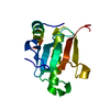
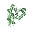
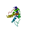
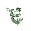
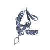
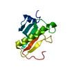
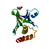
| Title | Structure of Cu(II)azurin with the metal-binding loop sequence "CTFPGHSALM" replaced with "CTPHPFM" | ||||||
 Components Components | Azurin | ||||||
 Keywords Keywords | ELECTRON TRANSPORT / Blue copper-binding protein / greek-key beta-barrel | ||||||
| Function / homology |  Function and homology information Function and homology informationtransition metal ion binding / periplasmic space / electron transfer activity / copper ion binding / zinc ion binding / identical protein binding / plasma membrane Similarity search - Function | ||||||
| Biological species |   Pseudomonas aeruginosa (bacteria) Pseudomonas aeruginosa (bacteria) | ||||||
| Method |  X-RAY DIFFRACTION / MOLECULAR REPLACEMENT / Resolution: 1.61 Å X-RAY DIFFRACTION / MOLECULAR REPLACEMENT / Resolution: 1.61 Å | ||||||
 Authors Authors | Banfield, M.J. | ||||||
 Citation Citation | Journal: Proc.Natl.Acad.Sci.Usa / Year: 2006 Title: Basic requirements for a metal-binding site in a protein: The influence of loop shortening on the cupredoxin azurin. Authors: Li, C. / Yanagisawa, S. / Martins, B.M. / Messerschmidt, A. / Banfield, M.J. / Dennison, C. #1: Journal: J.Mol.Biol. / Year: 1991Title: Crystal structure analysis of oxidized Pseudomonas aeruginosa azurin at pH 5.5 and pH 9.0. A pH-induced conformational transition involves a peptide bond flip Authors: Nar, H. / Messerschmidt, A. / Huber, R. / van de Kamp, M. / Canters, G.W. #2: Journal: J.Am.Chem.Soc. / Year: 2004 Title: Loop-contraction mutagenesis of type 1 copper sites Authors: Yanagisawa, S. / Dennison, C. #3: Journal: J.Mol.Biol. / Year: 1994Title: Crystal structure ananlysis and refinement at 2.15 A resolution of amicyanin, a type I blue copper protein, from Thiobacillus versutus Authors: Romero, A. / Nar, H. / Huber, R. / Messerschmidt, A. / Kalverda, A.P. / Canters, G.W. / Durley, R. / Mathews, F.S. | ||||||
| History |
| ||||||
| Remark 999 | SEQUENCE Amino acid sequence CTFPGHSALM, involving residues 132 to 141 in the amino acid database ...SEQUENCE Amino acid sequence CTFPGHSALM, involving residues 132 to 141 in the amino acid database was replaced with the sequence CTPHPM in the deposition |
- Structure visualization
| Structure viewer | Molecule: MolmilJmol/JSmol |
|---|
- Downloads & links
-Download
| PDBx/mmCIF format | 2fta.cif.gz | 119.8 KB | Display | PDBx/mmCIF format |
|---|---|---|---|---|
| PDB format | pdb2fta.ent.gz | 91.3 KB | Display | PDB format |
| PDBx/mmJSON format | 2fta.json.gz | Tree view | PDBx/mmJSON format | |
| Others |  Other downloads Other downloads |
-Validation report
| Arichive directory | https://data.pdbj.org/pub/pdb/validation_reports/ft/2ftaftp://data.pdbj.org/pub/pdb/validation_reports/ft/2fta | HTTPS FTP |
|---|
-Related structure data
| Related structure data |  2ft6C  2ft7C  2ft8C  4azuS C: citing same article ( S: Starting model for refinement |
|---|---|
| Similar structure data |

-Links
 PDBj
PDBj


- Assembly
| Deposited unit | 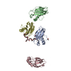
| ||||||||
|---|---|---|---|---|---|---|---|---|---|
| 1 | 
| ||||||||
| 2 | 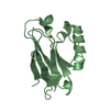
| ||||||||
| 3 | 
| ||||||||
| 4 | 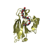
| ||||||||
| Unit cell |
| ||||||||
| Details | The biological assembly is monomeric (as found in the asymmetric unit) |
-Components
-Protein , 1 types, 4 molecules ABCD
| #1: Protein | Mass: 13730.549 Da / Num. of mol.: 4 Source method: isolated from a genetically manipulated source Source: (gene. exp.) Pseudomonas aeruginosa (bacteria) / Gene: azu / Plasmid: Trk99a / Production host: |
|---|
-Non-polymers , 5 types, 457 molecules 


| #2: Chemical | ChemComp-CU /  Mass: 63.546 Da / Num. of mol.: 4 / Source method: obtained synthetically / Formula: Cu Mass: 63.546 Da / Num. of mol.: 4 / Source method: obtained synthetically / Formula: Cu#3: Chemical |  Mass: 414.488 Da / Num. of mol.: 2 / Source method: obtained synthetically / Formula: C18H38O10 / Comment: precipitant*YM Mass: 414.488 Da / Num. of mol.: 2 / Source method: obtained synthetically / Formula: C18H38O10 / Comment: precipitant*YM#4: Chemical |  Mass: 46.068 Da / Num. of mol.: 3 / Source method: obtained synthetically / Formula: C2H6O Mass: 46.068 Da / Num. of mol.: 3 / Source method: obtained synthetically / Formula: C2H6O#5: Chemical | ChemComp-PEG / |  Mass: 106.120 Da / Num. of mol.: 1 / Source method: obtained synthetically / Formula: C4H10O3 Mass: 106.120 Da / Num. of mol.: 1 / Source method: obtained synthetically / Formula: C4H10O3#6: Water | ChemComp-HOH / | Mass: 18.015 Da / Num. of mol.: 447 / Source method: isolated from a natural source / Formula: H2O |
|---|
-Details
| Has protein modification | Y |
|---|
-Experimental details
-Experiment
| Experiment | Method: X-RAY DIFFRACTION / Number of used crystals: 1 |
|---|
- Sample preparation
| Crystal | Density Matthews: 1.99 Å3/Da / Density % sol: 38.34 % |
|---|---|
| Crystal grow | Temperature: 293 K / Method: vapor diffusion, hanging drop / pH: 7 Details: 0.1M Potassium Thiocyanate, 30% PEG MME 2000, pH 7, VAPOR DIFFUSION, HANGING DROP, temperature 293K |
-Data collection
| Diffraction | Mean temperature: 100 K |
|---|---|
| Diffraction source | Source: ROTATING ANODE / Type: ENRAF-NONIUS / Wavelength: 1.542 Å |
| Detector | Type: MARRESEARCH / Detector: IMAGE PLATE / Date: Oct 20, 2005 |
| Radiation | Protocol: SINGLE WAVELENGTH / Monochromatic (M) / Laue (L): M / Scattering type: x-ray |
| Radiation wavelength | Wavelength: 1.542 Å / Relative weight: 1 |
| Reflection | Resolution: 1.61→30 Å / Num. all: 55423 / Num. obs: 55423 / % possible obs: 99.4 % / Observed criterion σ(F): 0 / Observed criterion σ(I): 0 / Redundancy: 3.7 % / Biso Wilson estimate: 26.043 Å2 / Rmerge(I) obs: 0.039 / Rsym value: 0.039 / Net I/σ(I): 18.01 |
| Reflection shell | Resolution: 1.61→1.7 Å / % possible obs: 98.4 % / Rmerge(I) obs: 0.279 / Mean I/σ(I) obs: 4.4 / Num. measured obs: 24724 / Num. unique obs: 8203 / Rsym value: 0.279 / % possible all: 0.987 |
- Processing
| Software |
| ||||||||||||||||||||||||||||||||||||||||||||||||||||||||||||||||||||||||||||||||||||||||||||||||||||||||||||||||||||||||||||||||||||||||||||||||||||||||||||||||||||||||||||||||||||||||||||||||||||||||||||
|---|---|---|---|---|---|---|---|---|---|---|---|---|---|---|---|---|---|---|---|---|---|---|---|---|---|---|---|---|---|---|---|---|---|---|---|---|---|---|---|---|---|---|---|---|---|---|---|---|---|---|---|---|---|---|---|---|---|---|---|---|---|---|---|---|---|---|---|---|---|---|---|---|---|---|---|---|---|---|---|---|---|---|---|---|---|---|---|---|---|---|---|---|---|---|---|---|---|---|---|---|---|---|---|---|---|---|---|---|---|---|---|---|---|---|---|---|---|---|---|---|---|---|---|---|---|---|---|---|---|---|---|---|---|---|---|---|---|---|---|---|---|---|---|---|---|---|---|---|---|---|---|---|---|---|---|---|---|---|---|---|---|---|---|---|---|---|---|---|---|---|---|---|---|---|---|---|---|---|---|---|---|---|---|---|---|---|---|---|---|---|---|---|---|---|---|---|---|---|---|---|---|---|---|---|---|
| Refinement | Method to determine structure: MOLECULAR REPLACEMENT Starting model: PDB ENTRY 4AZU Resolution: 1.61→30 Å / FOM work R set: 0.789 / Isotropic thermal model: isotropic / Cross valid method: THROUGHOUT / σ(F): 0 / Stereochemistry target values: Engh & Huber
| ||||||||||||||||||||||||||||||||||||||||||||||||||||||||||||||||||||||||||||||||||||||||||||||||||||||||||||||||||||||||||||||||||||||||||||||||||||||||||||||||||||||||||||||||||||||||||||||||||||||||||||
| Displacement parameters | Biso mean: 25.978 Å2 | ||||||||||||||||||||||||||||||||||||||||||||||||||||||||||||||||||||||||||||||||||||||||||||||||||||||||||||||||||||||||||||||||||||||||||||||||||||||||||||||||||||||||||||||||||||||||||||||||||||||||||||
| Refine analyze |
| ||||||||||||||||||||||||||||||||||||||||||||||||||||||||||||||||||||||||||||||||||||||||||||||||||||||||||||||||||||||||||||||||||||||||||||||||||||||||||||||||||||||||||||||||||||||||||||||||||||||||||||
| Refinement step | Cycle: LAST / Resolution: 1.61→30 Å
| ||||||||||||||||||||||||||||||||||||||||||||||||||||||||||||||||||||||||||||||||||||||||||||||||||||||||||||||||||||||||||||||||||||||||||||||||||||||||||||||||||||||||||||||||||||||||||||||||||||||||||||
| Refine LS restraints |
| ||||||||||||||||||||||||||||||||||||||||||||||||||||||||||||||||||||||||||||||||||||||||||||||||||||||||||||||||||||||||||||||||||||||||||||||||||||||||||||||||||||||||||||||||||||||||||||||||||||||||||||
| LS refinement shell | Refine-ID: X-RAY DIFFRACTION / Total num. of bins used: 33
|
