 Movie
Movie Controller

[English] 日本語
 Yorodumi
- PDB-3ily: Apo crystal structure of protein tyrosine phosphatase from Entamo... -
+ Open data

- Basic information
| Entry | Database: PDB / ID: 3ily | ||||||
|---|---|---|---|---|---|---|---|
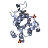
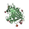
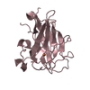
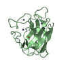
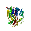
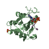
| Title | Apo crystal structure of protein tyrosine phosphatase from Entamoeba histolytica featuring a disordered active site | ||||||
 Components Components | Protein tyrosine phosphatase, putative | ||||||
 Keywords Keywords | HYDROLASE / NIAID / SSGCID / Seattle Structural Genomics Center for Infectious Disease / parasitic protozoan / dysentery | ||||||
| Function / homology |  Function and homology information Function and homology informationacid phosphatase / acid phosphatase activity / protein tyrosine phosphatase activity Similarity search - Function | ||||||
| Biological species |  Entamoeba histolytica HM-1:IMSS (eukaryote) Entamoeba histolytica HM-1:IMSS (eukaryote) | ||||||
| Method |  X-RAY DIFFRACTION / MOLECULAR REPLACEMENT / molecular replacement / Resolution: 2.2 Å X-RAY DIFFRACTION / MOLECULAR REPLACEMENT / molecular replacement / Resolution: 2.2 Å | ||||||
 Authors Authors | Seattle Structural Genomics Center for Infectious Disease (SSGCID) | ||||||
 Citation Citation | Journal: Mol.Biochem.Parasitol. / Year: 2014 Title: Crystal structure and putative substrate identification for the Entamoeba histolytica low molecular weight tyrosine phosphatase. Authors: Linford, A.S. / Jiang, N.M. / Edwards, T.E. / Sherman, N.E. / Van Voorhis, W.C. / Stewart, L.J. / Myler, P.J. / Staker, B.L. / Petri, W.A. | ||||||
| History |
|
- Structure visualization
| Structure viewer | Molecule: MolmilJmol/JSmol |
|---|
- Downloads & links
-Download
| PDBx/mmCIF format | 3ily.cif.gz | 70.2 KB | Display | PDBx/mmCIF format |
|---|---|---|---|---|
| PDB format | pdb3ily.ent.gz | 51.2 KB | Display | PDB format |
| PDBx/mmJSON format | 3ily.json.gz | Tree view | PDBx/mmJSON format | |
| Others |  Other downloads Other downloads |
-Validation report
| Arichive directory | https://data.pdbj.org/pub/pdb/validation_reports/il/3ilyftp://data.pdbj.org/pub/pdb/validation_reports/il/3ily | HTTPS FTP |
|---|
-Related structure data
| Related structure data | 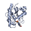 3idoSC  3js5C 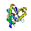 3jviC S: Starting model for refinement C: citing same article ( |
|---|---|
| Similar structure data | |
| Other databases |


-Links
 PDBj
PDBj
- Assembly
| Deposited unit | 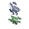
| ||||||||
|---|---|---|---|---|---|---|---|---|---|
| 1 | 
| ||||||||
| 2 | 
| ||||||||
| Unit cell |
|
-Components
| #1: Protein | Mass: 20382.250 Da / Num. of mol.: 2 Source method: isolated from a genetically manipulated source Source: (gene. exp.) Entamoeba histolytica HM-1:IMSS (eukaryote)Gene: EHI_153650 / Production host:  #2: Water | ChemComp-HOH / |  Mass: 18.015 Da / Num. of mol.: 70 / Source method: isolated from a natural source / Formula: H2O Mass: 18.015 Da / Num. of mol.: 70 / Source method: isolated from a natural source / Formula: H2O |
|---|
-Experimental details
-Experiment
| Experiment | Method: X-RAY DIFFRACTION / Number of used crystals: 1 |
|---|
- Sample preparation
| Crystal grow | Method: vapor diffusion, sitting drop Details: ProPlex Screen condition A9, 0.1 M MES pH 6.0, 20% PEG MME 2000, 0.2 M NaCl, 20% glycerol as cryo-protectant, 26.1 mg/mL protein, crystal tracking ID 203694a9, VAPOR DIFFUSION, SITTING DROP |
|---|
-Data collection
| Diffraction | Mean temperature: 100 K |
|---|---|
| Diffraction source | Source: ROTATING ANODE / Type: RIGAKU FR-E+ SUPERBRIGHT / Wavelength: 1.5418 Å |
| Detector | Type: RIGAKU SATURN 944+ / Detector: CCD / Date: Jul 30, 2009 |
| Radiation | Protocol: SINGLE WAVELENGTH / Monochromatic (M) / Laue (L): M / Scattering type: x-ray |
| Radiation wavelength | Wavelength: 1.5418 Å / Relative weight: 1 |
| Reflection | Resolution: 2.2→22.84 Å / Num. obs: 13504 / % possible obs: 98.3 % / Redundancy: 4.5 % / Rmerge(I) obs: 0.063 / Χ2: 1.042 / Net I/σ(I): 23.6 |
| Reflection shell | Resolution: 2.2→2.28 Å / Redundancy: 3.4 % / Rmerge(I) obs: 0.316 / Mean I/σ(I) obs: 4.3 / Num. unique all: 1154 / Χ2: 0.957 / % possible all: 85.4 |
-Phasing
| Phasing | Method: molecular replacement | |||||||||
|---|---|---|---|---|---|---|---|---|---|---|
| Phasing MR | Rfactor: 45.5 / Model details: Phaser MODE: MR_AUTO
|
- Processing
| Software |
| |||||||||||||||||||||||||||||||||||||||||||||||||||||||||||||||||
|---|---|---|---|---|---|---|---|---|---|---|---|---|---|---|---|---|---|---|---|---|---|---|---|---|---|---|---|---|---|---|---|---|---|---|---|---|---|---|---|---|---|---|---|---|---|---|---|---|---|---|---|---|---|---|---|---|---|---|---|---|---|---|---|---|---|---|
| Refinement | Method to determine structure: MOLECULAR REPLACEMENT Starting model: PDB enry 3IDO Resolution: 2.2→22.84 Å / Cor.coef. Fo:Fc: 0.948 / Cor.coef. Fo:Fc free: 0.931 / WRfactor Rfree: 0.262 / WRfactor Rwork: 0.207 / Occupancy max: 1 / Occupancy min: 0.5 / FOM work R set: 0.77 / SU B: 7.815 / SU ML: 0.198 / SU R Cruickshank DPI: 0.379 / SU Rfree: 0.253 / Cross valid method: THROUGHOUT / σ(F): 0 / ESU R: 0.379 / ESU R Free: 0.253 / Stereochemistry target values: MAXIMUM LIKELIHOOD Details: HYDROGENS HAVE BEEN ADDED IN THE RIDING POSITIONS U VALUES: REFINED INDIVIDUALLY
| |||||||||||||||||||||||||||||||||||||||||||||||||||||||||||||||||
| Solvent computation | Ion probe radii: 0.8 Å / Shrinkage radii: 0.8 Å / VDW probe radii: 1.4 Å / Solvent model: MASK | |||||||||||||||||||||||||||||||||||||||||||||||||||||||||||||||||
| Displacement parameters | Biso max: 73.12 Å2 / Biso mean: 37.906 Å2 / Biso min: 21.25 Å2
| |||||||||||||||||||||||||||||||||||||||||||||||||||||||||||||||||
| Refinement step | Cycle: LAST / Resolution: 2.2→22.84 Å
| |||||||||||||||||||||||||||||||||||||||||||||||||||||||||||||||||
| Refine LS restraints |
| |||||||||||||||||||||||||||||||||||||||||||||||||||||||||||||||||
| LS refinement shell | Resolution: 2.203→2.26 Å / Total num. of bins used: 20
|
