 Movie
Movie Controller

[English] 日本語
 Yorodumi
- PDB-1xtm: Crystal structure of the double mutant Y88H-P104H of a SOD-like p... -
+ Open data

- Basic information
| Entry | Database: PDB / ID: 1xtm | ||||||
|---|---|---|---|---|---|---|---|
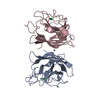
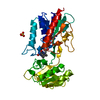
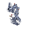
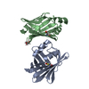
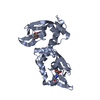
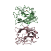
| Title | Crystal structure of the double mutant Y88H-P104H of a SOD-like protein from Bacillus subtilis. | ||||||
 Components Components | Hypothetical superoxide dismutase-like protein yojM | ||||||
 Keywords Keywords | STRUCTURAL GENOMICS / UNKNOWN FUNCTION / SOD / Cu-Zn SOD / SOD-like / superoxide dismutase mutants | ||||||
| Function / homology |  Function and homology information Function and homology informationsuperoxide dismutase activity / removal of superoxide radicals / copper ion binding / zinc ion binding / identical protein binding / plasma membrane Similarity search - Function | ||||||
| Biological species |  | ||||||
| Method |  X-RAY DIFFRACTION / SYNCHROTRON / MOLECULAR REPLACEMENT / Resolution: 1.6 Å X-RAY DIFFRACTION / SYNCHROTRON / MOLECULAR REPLACEMENT / Resolution: 1.6 Å | ||||||
 Authors Authors | Calderone, V. / Mangani, S. / Banci, L. / Benvenuti, M. / Bertini, I. / Fantoni, A. / Viezzoli, M.S. | ||||||
 Citation Citation | Journal: J.Am.Chem.Soc. / Year: 2005 Title: From an Inactive Prokaryotic SOD Homologue to an Active Protein through Site-Directed Mutagenesis. Authors: Banci, L. / Benvenuti, M. / Bertini, I. / Cabelli, D.E. / Calderone, V. / Fantoni, A. / Mangani, S. / Migliardi, M. / Viezzoli, M.S. #1: Journal: Proc.Natl.Acad.Sci.USA / Year: 2005Title: A prokaryotic superoxide dismutase paralog lacking two Cu ligands: from largely unstructured in solution to ordered in the crystal. Authors: Banci, L. / Bertini, I. / Calderone, V. / Cramaro, F. / Del Conte, R. / Fantoni, A. / Mangani, S. / Quattrone, A. / Viezzoli, M.S. | ||||||
| History |
|
- Structure visualization
| Structure viewer | Molecule: MolmilJmol/JSmol |
|---|
- Downloads & links
-Download
| PDBx/mmCIF format | 1xtm.cif.gz | 78.6 KB | Display | PDBx/mmCIF format |
|---|---|---|---|---|
| PDB format | pdb1xtm.ent.gz | 57.6 KB | Display | PDB format |
| PDBx/mmJSON format | 1xtm.json.gz | Tree view | PDBx/mmJSON format | |
| Others |  Other downloads Other downloads |
-Validation report
| Arichive directory | https://data.pdbj.org/pub/pdb/validation_reports/xt/1xtmftp://data.pdbj.org/pub/pdb/validation_reports/xt/1xtm | HTTPS FTP |
|---|
-Related structure data
| Related structure data | 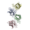 1xtlC 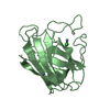 1s4iS S: Starting model for refinement C: citing same article ( |
|---|---|
| Similar structure data |


-Links
 PDBj
PDBj


- Assembly
| Deposited unit | 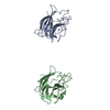
| ||||||||
|---|---|---|---|---|---|---|---|---|---|
| 1 | 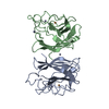
| ||||||||
| 2 | 
| ||||||||
| 3 | 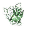
| ||||||||
| Unit cell |
| ||||||||
| Details | The protein is a monomer in vivo but there are two molecules. |
-Components
| #1: Protein | Mass: 18754.797 Da / Num. of mol.: 2 / Mutation: Y67H, P83H Source method: isolated from a genetically manipulated source Source: (gene. exp.) #2: Chemical |   Mass: 63.546 Da / Num. of mol.: 2 / Source method: obtained synthetically / Formula: Cu Mass: 63.546 Da / Num. of mol.: 2 / Source method: obtained synthetically / Formula: Cu#3: Chemical | ChemComp-ZN /   Mass: 65.409 Da / Num. of mol.: 5 / Source method: obtained synthetically / Formula: Zn Mass: 65.409 Da / Num. of mol.: 5 / Source method: obtained synthetically / Formula: Zn#4: Water | ChemComp-HOH / |  Mass: 18.015 Da / Num. of mol.: 226 / Source method: isolated from a natural source / Formula: H2O Mass: 18.015 Da / Num. of mol.: 226 / Source method: isolated from a natural source / Formula: H2OHas protein modification | Y | |
|---|
-Experimental details
-Experiment
| Experiment | Method: X-RAY DIFFRACTION / Number of used crystals: 1 |
|---|
- Sample preparation
| Crystal | Density Matthews: 2.14 Å3/Da / Density % sol: 42.59 % |
|---|---|
| Crystal grow | Temperature: 298 K / Method: vapor diffusion, sitting drop / pH: 4.6 Details: 0.1M Na Acetate, 30% PEG4000, 0.2M Ammonium Sulphate, 1mM ZnCl2, pH 4.6, VAPOR DIFFUSION, SITTING DROP, temperature 298K |
-Data collection
| Diffraction | Mean temperature: 100 K |
|---|---|
| Diffraction source | Source: SYNCHROTRON / Site: EMBL/DESY, HAMBURG  / Beamline: BW7A / Wavelength: 1.2768 Å / Beamline: BW7A / Wavelength: 1.2768 Å |
| Detector | Type: MARRESEARCH / Detector: CCD / Date: May 20, 2004 / Details: Double crystal focussing mono chromator |
| Radiation | Monochromator: Double crystal focussing mono chromator / Protocol: SINGLE WAVELENGTH / Monochromatic (M) / Laue (L): M / Scattering type: x-ray |
| Radiation wavelength | Wavelength: 1.2768 Å / Relative weight: 1 |
| Reflection | Resolution: 1.6→28.8 Å / Num. all: 43173 / Num. obs: 43173 / % possible obs: 99.6 % / Observed criterion σ(F): 0 / Observed criterion σ(I): 0 / Redundancy: 7 % / Biso Wilson estimate: 21.907 Å2 / Rmerge(I) obs: 0.057 / Rsym value: 0.057 / Net I/σ(I): 9.1 |
| Reflection shell | Resolution: 1.6→1.69 Å / Redundancy: 6.9 % / Rmerge(I) obs: 0.478 / Mean I/σ(I) obs: 1.6 / Num. unique all: 6245 / Rsym value: 0.478 / % possible all: 99.2 |
- Processing
| Software |
| ||||||||||||||||||||||||||||||||||||||||||||||||||||||||||||||||||||||||||||||||||||||||||||||||||||||||||||||||||||||||||||||||||||||||||||||||||||||||||||||||
|---|---|---|---|---|---|---|---|---|---|---|---|---|---|---|---|---|---|---|---|---|---|---|---|---|---|---|---|---|---|---|---|---|---|---|---|---|---|---|---|---|---|---|---|---|---|---|---|---|---|---|---|---|---|---|---|---|---|---|---|---|---|---|---|---|---|---|---|---|---|---|---|---|---|---|---|---|---|---|---|---|---|---|---|---|---|---|---|---|---|---|---|---|---|---|---|---|---|---|---|---|---|---|---|---|---|---|---|---|---|---|---|---|---|---|---|---|---|---|---|---|---|---|---|---|---|---|---|---|---|---|---|---|---|---|---|---|---|---|---|---|---|---|---|---|---|---|---|---|---|---|---|---|---|---|---|---|---|---|---|---|---|
| Refinement | Method to determine structure: MOLECULAR REPLACEMENT Starting model: 1S4I Resolution: 1.6→25.6 Å / Cor.coef. Fo:Fc: 0.928 / Cor.coef. Fo:Fc free: 0.919 / SU B: 2.43 / SU ML: 0.084 / Cross valid method: THROUGHOUT / σ(F): 0 / σ(I): 0 / ESU R: 0.121 / ESU R Free: 0.11 / Stereochemistry target values: MAXIMUM LIKELIHOOD
| ||||||||||||||||||||||||||||||||||||||||||||||||||||||||||||||||||||||||||||||||||||||||||||||||||||||||||||||||||||||||||||||||||||||||||||||||||||||||||||||||
| Solvent computation | Ion probe radii: 0.8 Å / Shrinkage radii: 0.8 Å / VDW probe radii: 1.2 Å / Solvent model: MASK | ||||||||||||||||||||||||||||||||||||||||||||||||||||||||||||||||||||||||||||||||||||||||||||||||||||||||||||||||||||||||||||||||||||||||||||||||||||||||||||||||
| Displacement parameters | Biso mean: 26.866 Å2
| ||||||||||||||||||||||||||||||||||||||||||||||||||||||||||||||||||||||||||||||||||||||||||||||||||||||||||||||||||||||||||||||||||||||||||||||||||||||||||||||||
| Refine analyze | Luzzati sigma a obs: 0.084 Å | ||||||||||||||||||||||||||||||||||||||||||||||||||||||||||||||||||||||||||||||||||||||||||||||||||||||||||||||||||||||||||||||||||||||||||||||||||||||||||||||||
| Refinement step | Cycle: LAST / Resolution: 1.6→25.6 Å
| ||||||||||||||||||||||||||||||||||||||||||||||||||||||||||||||||||||||||||||||||||||||||||||||||||||||||||||||||||||||||||||||||||||||||||||||||||||||||||||||||
| Refine LS restraints |
| ||||||||||||||||||||||||||||||||||||||||||||||||||||||||||||||||||||||||||||||||||||||||||||||||||||||||||||||||||||||||||||||||||||||||||||||||||||||||||||||||
| LS refinement shell | Resolution: 1.6→1.641 Å / Total num. of bins used: 20
|
