 Movie
Movie Controller


+ Open data

- Basic information
| Entry | Database: PDB / ID: 1vdc | ||||||
|---|---|---|---|---|---|---|---|
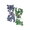
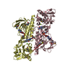
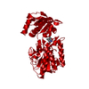
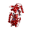
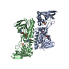
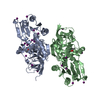
| Title | STRUCTURE OF NADPH DEPENDENT THIOREDOXIN REDUCTASE | ||||||
 Components Components | NADPH DEPENDENT THIOREDOXIN REDUCTASE | ||||||
 Keywords Keywords | OXIDOREDUCTASE / HYPOTHETICAL PROTEIN / REDOX-ACTIVE CENTER / DISULFIDE OXIDOREDUCTASE / THIOREDOXIN REDUCTASE / FLAVIN ADENINE DINULEOTIDE | ||||||
| Function / homology |  Function and homology information Function and homology informationthioredoxin-disulfide reductase (NADPH) / chloroplast envelope / thioredoxin-disulfide reductase (NADPH) activity / positive regulation of cell division / removal of superoxide radicals / cell redox homeostasis / mitochondrion / plasma membrane / cytosol Similarity search - Function | ||||||
| Biological species |  | ||||||
| Method |  X-RAY DIFFRACTION / SYNCHROTRON / MOLECULAR REPLACEMENT / Resolution: 2.5 Å X-RAY DIFFRACTION / SYNCHROTRON / MOLECULAR REPLACEMENT / Resolution: 2.5 Å | ||||||
 Authors Authors | Dai, S. / Eklund, H. | ||||||
 Citation Citation | Journal: J.Mol.Biol. / Year: 1996 Title: Crystal structure of Arabidopsis thaliana NADPH dependent thioredoxin reductase at 2.5 A resolution. Authors: Dai, S. / Saarinen, M. / Ramaswamy, S. / Meyer, Y. / Jacquot, J.P. / Eklund, H. #1: Journal: J.Mol.Biol. / Year: 1994Title: Crystal Structure of Escherichia Coli Thioredoxin Reductase Refined at 2 A Resolution. Implications for a Large Conformational Change During Catalysis Authors: Waksman, G. / Krishna, T.S. / Williams Junior, C.H. / Kuriyan, J. | ||||||
| History |
|
- Structure visualization
| Structure viewer | Molecule: MolmilJmol/JSmol |
|---|
- Downloads & links
-Download
| PDBx/mmCIF format | 1vdc.cif.gz | 84.9 KB | Display | PDBx/mmCIF format |
|---|---|---|---|---|
| PDB format | pdb1vdc.ent.gz | 62 KB | Display | PDB format |
| PDBx/mmJSON format | 1vdc.json.gz | Tree view | PDBx/mmJSON format | |
| Others |  Other downloads Other downloads |
-Validation report
| Arichive directory | https://data.pdbj.org/pub/pdb/validation_reports/vd/1vdcftp://data.pdbj.org/pub/pdb/validation_reports/vd/1vdc | HTTPS FTP |
|---|
-Related structure data
| Related structure data |  1trbS S: Starting model for refinement |
|---|---|
| Similar structure data |
-Links
 PDBj
PDBj


- Assembly
| Deposited unit | 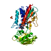
| ||||||||
|---|---|---|---|---|---|---|---|---|---|
| 1 | 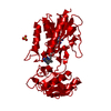
| ||||||||
| Unit cell |
|
-Components
| #1: Protein | Mass: 35420.055 Da / Num. of mol.: 1 Source method: isolated from a genetically manipulated source Source: (gene. exp.)  | ||
|---|---|---|---|
| #2: Chemical | ChemComp-SO4 /   Mass: 96.063 Da / Num. of mol.: 1 / Source method: obtained synthetically / Formula: SO4 Mass: 96.063 Da / Num. of mol.: 1 / Source method: obtained synthetically / Formula: SO4 | ||
| #3: Chemical | ChemComp-FAD /   Mass: 785.550 Da / Num. of mol.: 1 / Source method: obtained synthetically / Formula: C27H33N9O15P2 / Comment: FAD*YM Mass: 785.550 Da / Num. of mol.: 1 / Source method: obtained synthetically / Formula: C27H33N9O15P2 / Comment: FAD*YM | ||
| #4: Water | ChemComp-HOH /  Mass: 18.015 Da / Num. of mol.: 326 / Source method: isolated from a natural source / Formula: H2O Mass: 18.015 Da / Num. of mol.: 326 / Source method: isolated from a natural source / Formula: H2O | ||
| Has protein modification | Y | ||
| Nonpolymer details | NADPH CAN ALSO BE SEEN WITH LOW OCCUPANCY BUT NO COORDINATE| Sequence details | PROTEIN SEQUENCE NUMBERING IS BASED ON E. COLI NTR SEQUENCE AND IS NOT THE SAME AS THE SEQUENCE ...PROTEIN SEQUENCE NUMBERING IS BASED ON E. COLI NTR SEQUENCE AND IS NOT THE SAME AS THE SEQUENCE NUMBERING IN GENBANK. | |
-Experimental details
-Experiment
| Experiment | Method: X-RAY DIFFRACTION / Number of used crystals: 2 |
|---|
- Sample preparation
| Crystal | Density Matthews: 5.05 Å3/Da / Density % sol: 70.6 % | ||||||||||||||||||
|---|---|---|---|---|---|---|---|---|---|---|---|---|---|---|---|---|---|---|---|
| Crystal grow | Details: 7% ISOPROPANOL (OR 4% MPD) AND 1.8 M AMMONIUM SULFATE | ||||||||||||||||||
| Crystal grow | *PLUS Method: vapor diffusion, hanging drop | ||||||||||||||||||
| Components of the solutions | *PLUS
|
-Data collection
| Diffraction | Mean temperature: 175 K |
|---|---|
| Diffraction source | Source: SYNCHROTRON / Site: EMBL/DESY, HAMBURG  / Type: EMBL/DESY, HAMBURG / Wavelength: 0.857 / Type: EMBL/DESY, HAMBURG / Wavelength: 0.857 |
| Detector | Type: RIGAKU / Detector: IMAGE PLATE / Date: Jul 1, 1995 |
| Radiation | Monochromatic (M) / Laue (L): M / Scattering type: x-ray |
| Radiation wavelength | Wavelength: 0.857 Å / Relative weight: 1 |
| Reflection | Resolution: 2.5→20 Å / Num. obs: 25745 / % possible obs: 100 % / Observed criterion σ(I): 0 / Redundancy: 6.81 % / Biso Wilson estimate: 30.92 Å2 / Rmerge(I) obs: 0.085 / Net I/σ(I): 7.7 |
| Reflection shell | Resolution: 2.5→2.54 Å / Rmerge(I) obs: 0.487 / % possible all: 100 |
| Reflection | *PLUS Num. obs: 20556 / Num. measured all: 139995 |
| Reflection shell | *PLUS % possible obs: 100 % |
- Processing
| Software |
| ||||||||||||||||||||||||||||||||||||||||||||||||||||||||||||
|---|---|---|---|---|---|---|---|---|---|---|---|---|---|---|---|---|---|---|---|---|---|---|---|---|---|---|---|---|---|---|---|---|---|---|---|---|---|---|---|---|---|---|---|---|---|---|---|---|---|---|---|---|---|---|---|---|---|---|---|---|---|
| Refinement | Method to determine structure: MOLECULAR REPLACEMENT Starting model: PDB ENTRY 1TRB Resolution: 2.5→20 Å / σ(F): 1
| ||||||||||||||||||||||||||||||||||||||||||||||||||||||||||||
| Displacement parameters | Biso mean: 31.6 Å2 | ||||||||||||||||||||||||||||||||||||||||||||||||||||||||||||
| Refinement step | Cycle: LAST / Resolution: 2.5→20 Å
| ||||||||||||||||||||||||||||||||||||||||||||||||||||||||||||
| Refine LS restraints |
| ||||||||||||||||||||||||||||||||||||||||||||||||||||||||||||
| LS refinement shell | Resolution: 2.5→2.61 Å
| ||||||||||||||||||||||||||||||||||||||||||||||||||||||||||||
| Software | *PLUS Name: X-PLOR / Classification: refinement | ||||||||||||||||||||||||||||||||||||||||||||||||||||||||||||
| Refinement | *PLUS | ||||||||||||||||||||||||||||||||||||||||||||||||||||||||||||
| Solvent computation | *PLUS | ||||||||||||||||||||||||||||||||||||||||||||||||||||||||||||
| Displacement parameters | *PLUS | ||||||||||||||||||||||||||||||||||||||||||||||||||||||||||||
| Refine LS restraints | *PLUS
|
