 Movie
Movie Controller

[English] 日本語
 Yorodumi
- PDB-1cl0: CRYSTAL STRUCTURE OF REDUCED THIOREDOXIN REDUCTASE FROM ESCHERICH... -
+ Open data

- Basic information
| Entry | Database: PDB / ID: 1cl0 | ||||||
|---|---|---|---|---|---|---|---|
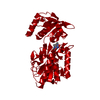
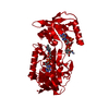
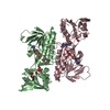
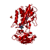
| Title | CRYSTAL STRUCTURE OF REDUCED THIOREDOXIN REDUCTASE FROM ESCHERICHIA COLI. | ||||||
 Components Components | THIOREDOXIN REDUCTASE | ||||||
 Keywords Keywords | OXIDOREDUCTASE / FLAVOENZYME | ||||||
| Function / homology |  Function and homology information Function and homology informationthioredoxin-disulfide reductase complex / thioredoxin-disulfide reductase (NADPH) / thioredoxin-disulfide reductase (NADPH) activity / removal of superoxide radicals / cell redox homeostasis / flavin adenine dinucleotide binding / cytosol Similarity search - Function | ||||||
| Biological species |  | ||||||
| Method |  X-RAY DIFFRACTION / MOLECULAR REPLACEMENT / Resolution: 2.5 Å X-RAY DIFFRACTION / MOLECULAR REPLACEMENT / Resolution: 2.5 Å | ||||||
 Authors Authors | Lennon, B.W. / Williams Jr, C.H. / Ludwig, M.L. | ||||||
 Citation Citation | Journal: Protein Sci. / Year: 1999 Title: Crystal structure of reduced thioredoxin reductase from Escherichia coli: structural flexibility in the isoalloxazine ring of the flavin adenine dinucleotide cofactor. Authors: Lennon, B.W. / Williams Jr., C.H. / Ludwig, M.L. #1: Journal: J.Mol.Biol. / Year: 1994Title: Crystal structure of Escherichia coli thioredoxin reductase refined at 2 A resolution. Implications for a large conformational change during catalysis. Authors: Waksman, G. / Krishna, T.S. / Williams Jr., C.H. / Kuriyan, J. | ||||||
| History |
|
- Structure visualization
| Structure viewer | Molecule: MolmilJmol/JSmol |
|---|
- Downloads & links
-Download
| PDBx/mmCIF format | 1cl0.cif.gz | 78.3 KB | Display | PDBx/mmCIF format |
|---|---|---|---|---|
| PDB format | pdb1cl0.ent.gz | 57.3 KB | Display | PDB format |
| PDBx/mmJSON format | 1cl0.json.gz | Tree view | PDBx/mmJSON format | |
| Others |  Other downloads Other downloads |
-Validation report
| Arichive directory | https://data.pdbj.org/pub/pdb/validation_reports/cl/1cl0ftp://data.pdbj.org/pub/pdb/validation_reports/cl/1cl0 | HTTPS FTP |
|---|
-Related structure data
| Related structure data | 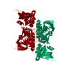 1tdeS S: Starting model for refinement |
|---|---|
| Similar structure data |

-Links
 PDBj
PDBj


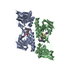
- Assembly
| Deposited unit | 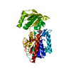
| ||||||||
|---|---|---|---|---|---|---|---|---|---|
| 1 | 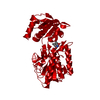
| ||||||||
| Unit cell |
|
-Components
| #1: Protein | Mass: 34529.766 Da / Num. of mol.: 1 Source method: isolated from a genetically manipulated source Details: FAD AND ACTIVE SITE DISULFIDE (CYS 135 AND CYS 138) REDUCED BY SODIUM DITHIONITE AFTER CRYSTALLIZATION Source: (gene. exp.) |
|---|---|
| #2: Chemical | ChemComp-FAD /   Mass: 785.550 Da / Num. of mol.: 1 / Source method: obtained synthetically / Formula: C27H33N9O15P2 / Comment: FAD*YM Mass: 785.550 Da / Num. of mol.: 1 / Source method: obtained synthetically / Formula: C27H33N9O15P2 / Comment: FAD*YM |
| #3: Water | ChemComp-HOH /  Mass: 18.015 Da / Num. of mol.: 109 / Source method: isolated from a natural source / Formula: H2O Mass: 18.015 Da / Num. of mol.: 109 / Source method: isolated from a natural source / Formula: H2O |
-Experimental details
-Experiment
| Experiment | Method: X-RAY DIFFRACTION / Number of used crystals: 1 |
|---|
- Sample preparation
| Crystal | Density Matthews: 2.45 Å3/Da / Density % sol: 50.8 % | ||||||||||||||||||||||||||||||
|---|---|---|---|---|---|---|---|---|---|---|---|---|---|---|---|---|---|---|---|---|---|---|---|---|---|---|---|---|---|---|---|
| Crystal grow | pH: 8 / Details: pH 8.0 | ||||||||||||||||||||||||||||||
| Crystal grow | *PLUS Temperature: 25 ℃ / pH: 7 / Method: vapor diffusion, hanging drop | ||||||||||||||||||||||||||||||
| Components of the solutions | *PLUS
|
-Data collection
| Diffraction | Mean temperature: 113 K |
|---|---|
| Diffraction source | Source: ROTATING ANODE / Type: RIGAKU RUH3R / Wavelength: 1.5418 |
| Detector | Type: RIGAKU / Detector: IMAGE PLATE / Date: Mar 1, 1998 / Details: MIRRORS |
| Radiation | Monochromator: NI FILTER / Protocol: SINGLE WAVELENGTH / Monochromatic (M) / Laue (L): M / Scattering type: x-ray |
| Radiation wavelength | Wavelength: 1.5418 Å / Relative weight: 1 |
| Reflection | Resolution: 2.5→10 Å / Num. obs: 12009 / % possible obs: 96.5 % / Redundancy: 7.8 % / Biso Wilson estimate: 21.1 Å2 / Rsym value: 0.119 / Net I/σ(I): 9.5 |
| Reflection shell | Resolution: 2.5→2.65 Å / Mean I/σ(I) obs: 2.1 / % possible all: 82.4 |
| Reflection | *PLUS Rmerge(I) obs: 0.119 |
| Reflection shell | *PLUS % possible obs: 82.4 % |
- Processing
| Software |
| ||||||||||||||||||||||||||||||||||||||||||||||||||||||||||||
|---|---|---|---|---|---|---|---|---|---|---|---|---|---|---|---|---|---|---|---|---|---|---|---|---|---|---|---|---|---|---|---|---|---|---|---|---|---|---|---|---|---|---|---|---|---|---|---|---|---|---|---|---|---|---|---|---|---|---|---|---|---|
| Refinement | Method to determine structure: MOLECULAR REPLACEMENT Starting model: 1TDE Resolution: 2.5→10 Å / Rfactor Rfree error: 0.011 / Data cutoff high absF: 1000000 / Data cutoff low absF: 0.001 / Isotropic thermal model: GROUP / Cross valid method: THROUGHOUT / σ(F): 0 Details: BULK SOLVENT MODEL USED DISORDERED REGION (RESIDUES 225-230) WAS MODELED STEREOCHEMICALLY. A HYDROGEN BOND PROBABLY EXISTS BETWEEN THE GAMMA SULFURS OF RESIDUES CYS 135 AND CYS 138.
| ||||||||||||||||||||||||||||||||||||||||||||||||||||||||||||
| Displacement parameters | Biso mean: 24.4 Å2 | ||||||||||||||||||||||||||||||||||||||||||||||||||||||||||||
| Refine analyze |
| ||||||||||||||||||||||||||||||||||||||||||||||||||||||||||||
| Refinement step | Cycle: LAST / Resolution: 2.5→10 Å
| ||||||||||||||||||||||||||||||||||||||||||||||||||||||||||||
| Refine LS restraints |
| ||||||||||||||||||||||||||||||||||||||||||||||||||||||||||||
| LS refinement shell | Resolution: 2.5→2.65 Å / Rfactor Rfree error: 0.037 / Total num. of bins used: 6
| ||||||||||||||||||||||||||||||||||||||||||||||||||||||||||||
| Xplor file |
| ||||||||||||||||||||||||||||||||||||||||||||||||||||||||||||
| Software | *PLUS Name: X-PLOR / Version: 3.851 / Classification: refinement | ||||||||||||||||||||||||||||||||||||||||||||||||||||||||||||
| Refine LS restraints | *PLUS
|
