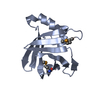
Resolution: 2.1→39.732 Å / SU ML: 0.3 / σ(F): 1.35 / Phase error: 20.02 / Stereochemistry target values: ML Details: A LOOP FROM RESIDUES 381-387 WAS DISORDERED IN THE STRUCTURE AND WAS THEREFORE NOT MODELLED.
Rfactor
Num. reflection
% reflection
Rfree
0.2131
483
4.7 %
Rwork
0.1821
-
-
obs
0.1835
10323
94.83 %
Solvent computation
Shrinkage radii: 0.95 Å / VDW probe radii: 1.2 Å / Solvent model: FLAT BULK SOLVENT MODEL / Bsol: 54.389 Å2 / ksol: 0.382 e/Å3
Displacement parameters
Biso mean: 42.4 Å2
Baniso -1
Baniso -2
Baniso -3
1-
-2.6955 Å2
0 Å2
0 Å2
2-
-
14.2707 Å2
0 Å2
3-
-
-
-11.5752 Å2
Refinement step
Cycle: LAST / Resolution: 2.1→39.732 Å
Protein
Nucleic acid
Ligand
Solvent
Total
Num. atoms
977
0
6
49
1032
Refine LS restraints
Refine-ID
Type
Dev ideal
Number
X-RAY DIFFRACTION
f_bond_d
0.008
1011
X-RAY DIFFRACTION
f_angle_d
1.011
1356
X-RAY DIFFRACTION
f_dihedral_angle_d
14.77
388
X-RAY DIFFRACTION
f_chiral_restr
0.068
145
X-RAY DIFFRACTION
f_plane_restr
0.005
169
LS refinement shell
Resolution (Å)
Rfactor Rfree
Num. reflection Rfree
Rfactor Rwork
Num. reflection Rwork
Refine-ID
% reflection obs (%)
2.1003-2.4041
0.2266
169
0.1988
3213
X-RAY DIFFRACTION
95
2.4041-3.0288
0.2284
161
0.1828
3256
X-RAY DIFFRACTION
95
3.0288-39.7393
0.203
153
0.178
3371
X-RAY DIFFRACTION
95
Refinement TLS params.
Method: refined / Refine-ID: X-RAY DIFFRACTION
ID
L11 (°2)
L12 (°2)
L13 (°2)
L22 (°2)
L23 (°2)
L33 (°2)
S11 (Å °)
S12 (Å °)
S13 (Å °)
S21 (Å °)
S22 (Å °)
S23 (Å °)
S31 (Å °)
S32 (Å °)
S33 (Å °)
T11 (Å2)
T12 (Å2)
T13 (Å2)
T22 (Å2)
T23 (Å2)
T33 (Å2)
Origin x (Å)
Origin y (Å)
Origin z (Å)
1
4.1069
1.3925
0.0424
0.6937
1.047
6.5276
-0.4317
-0.3151
0.3691
0.1144
0.023
0.2141
-0.6568
-1.081
0.5109
0.2492
0.0844
-0.046
0.2654
-0.0671
0.3207
-5.8275
-13.4135
-16.6145
2
8.1204
-4.0778
1.4552
5.9013
-3.8921
2.8824
0.3538
-0.2295
-1.8316
-0.3716
-0.9264
0.0664
2.111
-0.5337
0.3366
0.6782
-0.2188
0.1663
0.4543
0.075
0.7415
-6.3319
-27.4055
-15.4295
3
4.8852
0.7174
0.0681
1.4696
0.2347
4.3218
-0.0039
-0.1315
0.2546
0.2094
-0.2403
0.4932
-0.1494
-1.9324
0.1017
0.2293
0.0568
0.0766
0.6514
-0.0541
0.3734
-12.8374
-14.5609
-14.2879
4
6.3339
1.6919
-3.157
1.9312
-0.5674
8.1702
0.0343
-0.6403
-0.0606
0.3374
-0.1453
-0.0129
0.8145
0.2764
0.3765
0.3187
-0.0057
0.0331
0.2105
0.0385
0.2308
0.9335
-21.2398
-6.4586
5
4.6543
1.2119
-0.965
2.5593
-2.4312
3.0411
-0.6402
-0.3547
-1.1004
0.1143
-0.4228
-0.5651
1.3049
-0.4451
0.7014
0.4501
-0.078
0.0888
0.2877
0.0064
0.442
-2.9206
-24.5193
-8.2451
6
3.4124
0.3504
-0.1293
5.3719
1.8472
7.7126
-0.0267
-0.0923
0.3799
0.1798
-0.1944
-0.0575
-0.6474
0.0938
0.1437
0.2319
0.0174
-0.032
0.1491
0.0059
0.2474
2.8426
-9.3157
-9.2299
7
6.0179
0.5997
-5.0247
5.318
1.494
4.9306
0.2595
0.3609
0.2085
-0.4409
0.4111
-1.1615
0.3015
2.2999
0.0551
0.1468
-0.014
0.0449
1.1089
-0.211
0.8597
15.4018
-12.9522
-14.9308
Refinement TLS group
ID
Refine-ID
Refine TLS-ID
Selection details
1
X-RAY DIFFRACTION
1
(CHAINAANDRESID370:378)
2
X-RAY DIFFRACTION
2
(CHAINAANDRESID379:390)
3
X-RAY DIFFRACTION
3
(CHAINAANDRESID391:418)
4
X-RAY DIFFRACTION
4
(CHAINAANDRESID419:447)
5
X-RAY DIFFRACTION
5
(CHAINAANDRESID448:454)
6
X-RAY DIFFRACTION
6
(CHAINAANDRESID455:489)
7
X-RAY DIFFRACTION
7
(CHAINAANDRESID490:497)
+
About Yorodumi
-
News
-
Feb 9, 2022. New format data for meta-information of EMDB entries
New format data for meta-information of EMDB entries
Version 3 of the EMDB header file is now the official format.
The previous official version 1.9 will be removed from the archive.
In the structure databanks used in Yorodumi, some data are registered as the other names, "COVID-19 virus" and "2019-nCoV". Here are the details of the virus and the list of structure data.
Jan 31, 2019. EMDB accession codes are about to change! (news from PDBe EMDB page)
EMDB accession codes are about to change! (news from PDBe EMDB page)
The allocation of 4 digits for EMDB accession codes will soon come to an end. Whilst these codes will remain in use, new EMDB accession codes will include an additional digit and will expand incrementally as the available range of codes is exhausted. The current 4-digit format prefixed with “EMD-” (i.e. EMD-XXXX) will advance to a 5-digit format (i.e. EMD-XXXXX), and so on. It is currently estimated that the 4-digit codes will be depleted around Spring 2019, at which point the 5-digit format will come into force.
The EM Navigator/Yorodumi systems omit the EMD- prefix.
Related info.:Q: What is EMD? / ID/Accession-code notation in Yorodumi/EM Navigator
Yorodumi is a browser for structure data from EMDB, PDB, SASBDB, etc.
This page is also the successor to EM Navigator detail page, and also detail information page/front-end page for Omokage search.
The word "yorodu" (or yorozu) is an old Japanese word meaning "ten thousand". "mi" (miru) is to see.
Related info.:EMDB / PDB / SASBDB / Comparison of 3 databanks / Yorodumi Search / Aug 31, 2016. New EM Navigator & Yorodumi / Yorodumi Papers / Jmol/JSmol / Function and homology information / Changes in new EM Navigator and Yorodumi
 Movie
Movie Controller

 Yorodumi
 Open data
 Basic information
 Components
 Keywords
 Function and homology information
 X-RAY DIFFRACTION /
  Authors
 Citation
 Structure visualization
 Downloads & links
 Other downloads


 PDBj
PDBj

 Assembly


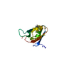
 Mass: 92.094 Da / Num. of mol.: 1 / Source method: obtained synthetically / Formula: C3H8O3
 Mass: 18.015 Da / Num. of mol.: 49 / Source method: isolated from a natural source / Formula: H2O
 Sample preparation
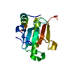
 / Beamline: I03 / Wavelength: 0.9745
  Processing