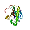
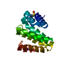
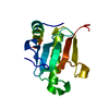
Mass: 14879.056 Da / Num. of mol.: 2 Source method: isolated from a genetically manipulated source Source: (gene. exp.) Bacteroides eggerthii DSM 20697 (bacteria) Gene: BACEGG_01585 / Plasmid: SpeedET / Production host: Escherichia Coli (E. coli) / Strain (production host): PB1 / References: UniProt: B7AGQ7
Has protein modification
Y
Sequence details
THE CONSTRUCT (25-150) WAS EXPRESSED WITH A PURIFICATION TAG MGSDKIHHHHHHENLYFQG. THE TAG WAS ...THE CONSTRUCT (25-150) WAS EXPRESSED WITH A PURIFICATION TAG MGSDKIHHHHHHENLYFQG. THE TAG WAS REMOVED WITH TEV PROTEASE LEAVING ONLY A GLYCINE (0) FOLLOWED BY THE TARGET SEQUENCE.
-
Experimental details
-
Experiment
Experiment
Method: X-RAY DIFFRACTION / Number of used crystals: 1
-
Sample preparation
Crystal
Density Matthews: 2.08 Å3/Da / Density % sol: 40.89 %
Type: DECTRIS PILATUS 6M / Detector: PIXEL / Date: May 23, 2013 Details: Flat mirror (vertical focusing); single crystal Si(111) bent monochromator (horizontal focusing)
Radiation
Monochromator: single crystal Si(111) bent / Protocol: MAD / Monochromatic (M) / Laue (L): M / Scattering type: x-ray
Radiation wavelength
ID
Wavelength (Å)
Relative weight
1
0.97839
1
2
0.91837
1
3
0.9798
1
Reflection
Resolution: 2.6→42.712 Å / Num. obs: 8129 / % possible obs: 93.4 % / Observed criterion σ(I): -3 / Redundancy: 5.033 % / Biso Wilson estimate: 91.209 Å2 / Rmerge F obs: 0.999 / Rmerge(I) obs: 0.043 / Rrim(I) all: 0.047 / Net I/σ(I): 20.71 / Num. measured all: 73713
Reflection shell
Resolution (Å)
Rmerge F obs
Rmerge(I) obs
Mean I/σ(I) obs
Num. measured obs
Num. possible
Num. unique obs
Rrim(I) all
Diffraction-ID
% possible all
2.55-2.64
0.708
1.054
1.2
2885
1543
584
1.175
1
37.8
2.64-2.75
0.829
0.758
1.9
8091
1633
1619
0.846
99.1
2.75-2.87
0.918
0.469
3.2
7146
1483
1466
0.525
98.9
2.87-3.02
0.977
0.233
5.9
8326
1574
1570
0.258
99.7
3.02-3.21
0.989
0.149
9.8
8116
1567
1563
0.166
99.7
3.21-3.46
0.997
0.087
15.5
7540
1589
1578
0.097
99.3
3.46-3.8
0.998
0.053
25.1
7960
1544
1537
0.059
99.5
3.8-4.35
0.999
0.033
36.8
7943
1581
1571
0.037
99.4
4.35-5.46
0.999
0.029
45.3
7902
1559
1556
0.032
99.8
5.46-42.712
0.999
0.026
49.3
7804
1614
1603
0.028
99.3
-
Phasing
Phasing
Method: MAD
-
Processing
Software
Name
Version
Classification
PDB_EXTRACT
3.1
dataextraction
XDS
datascaling
XSCALE
January10, 2014BUILT=20140307
datascaling
BUSTER-TNT
2.10.0
refinement
SHELX
phasing
SHARP
phasing
BUSTER
2.10.0
refinement
SHELXD
phasing
Refinement
Method to determine structure: MAD / Resolution: 2.6→42.712 Å / Cor.coef. Fo:Fc: 0.909 / Cor.coef. Fo:Fc free: 0.8535 / Occupancy max: 1 / Occupancy min: 0.75 / Cross valid method: THROUGHOUT / σ(F): 0 Details: 1. A MET-INHIBITION PROTOCOL WAS USED FOR SELENOMETHIONINE INCORPORATION DURING PROTEIN EXPRESSION. THE OCCUPANCY OF THE SE ATOMS IN THE MSE RESIDUES WAS REDUCED TO 0.75 FOR THE REDUCED ...Details: 1. A MET-INHIBITION PROTOCOL WAS USED FOR SELENOMETHIONINE INCORPORATION DURING PROTEIN EXPRESSION. THE OCCUPANCY OF THE SE ATOMS IN THE MSE RESIDUES WAS REDUCED TO 0.75 FOR THE REDUCED SCATTERING POWER DUE TO PARTIAL S-MET INCORPORATION. 2. ATOM RECORD CONTAINS SUM OF TLS AND RESIDUAL B FACTORS. ANISOU RECORD CONTAINS SUM OF TLS AND RESIDUAL U FACTORS. 3. THE MAD PHASES WERE USED AS RESTRAINTS DURING REFINEMENT.
In the structure databanks used in Yorodumi, some data are registered as the other names, "COVID-19 virus" and "2019-nCoV". Here are the details of the virus and the list of structure data.
Jan 31, 2019. EMDB accession codes are about to change! (news from PDBe EMDB page)
EMDB accession codes are about to change! (news from PDBe EMDB page)
The allocation of 4 digits for EMDB accession codes will soon come to an end. Whilst these codes will remain in use, new EMDB accession codes will include an additional digit and will expand incrementally as the available range of codes is exhausted. The current 4-digit format prefixed with “EMD-” (i.e. EMD-XXXX) will advance to a 5-digit format (i.e. EMD-XXXXX), and so on. It is currently estimated that the 4-digit codes will be depleted around Spring 2019, at which point the 5-digit format will come into force.
The EM Navigator/Yorodumi systems omit the EMD- prefix.
Related info.:Q: What is EMD? / ID/Accession-code notation in Yorodumi/EM Navigator
Yorodumi is a browser for structure data from EMDB, PDB, SASBDB, etc.
This page is also the successor to EM Navigator detail page, and also detail information page/front-end page for Omokage search.
The word "yorodu" (or yorozu) is an old Japanese word meaning "ten thousand". "mi" (miru) is to see.
Related info.:EMDB / PDB / SASBDB / Comparison of 3 databanks / Yorodumi Search / Aug 31, 2016. New EM Navigator & Yorodumi / Yorodumi Papers / Jmol/JSmol / Function and homology information / Changes in new EM Navigator and Yorodumi
 Movie
Movie Controller

 Yorodumi
 Open data
 Basic information
 Components
 Keywords
 Function and homology information
 Bacteroides eggerthii DSM 20697 (bacteria)
 X-RAY DIFFRACTION /
  Authors
 Citation
 Structure visualization
 Downloads & links
 Other downloads
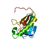


 PDBj
PDBj Assembly
 Sample preparation
 / Beamline: BL11-1 / Wavelength: 0.97839,0.91837,0.9798
 Processing