 Movie
Movie Controller

[English] 日本語
 Yorodumi
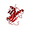
- PDB-3o1x: High resolution crystal structure of histidine triad nucleotide-b... -
+ Open data

- Basic information
| Entry | Database: PDB / ID: 3o1x | ||||||
|---|---|---|---|---|---|---|---|
| Title | High resolution crystal structure of histidine triad nucleotide-binding protein 1 (Hint1) C84A mutant from rabbit complexed with adenosine | ||||||
 Components Components | Histidine triad nucleotide-binding protein 1 | ||||||
 Keywords Keywords | HYDROLASE / HINT PROTEIN / HIT PROTEIN / ADENOSINE 5'-MONOPHOSPHORAMIDASE | ||||||
| Function / homology |  Function and homology information Function and homology informationpurine ribonucleotide catabolic process / Hydrolases; Acting on phosphorus-nitrogen bonds / adenosine 5'-monophosphoramidase activity / deSUMOylase activity / protein desumoylation / histone deacetylase complex / intrinsic apoptotic signaling pathway by p53 class mediator / Hydrolases; Acting on peptide bonds (peptidases); Cysteine endopeptidases / nucleotide binding / hydrolase activity ...purine ribonucleotide catabolic process / Hydrolases; Acting on phosphorus-nitrogen bonds / adenosine 5'-monophosphoramidase activity / deSUMOylase activity / protein desumoylation / histone deacetylase complex / intrinsic apoptotic signaling pathway by p53 class mediator / Hydrolases; Acting on peptide bonds (peptidases); Cysteine endopeptidases / nucleotide binding / hydrolase activity / regulation of DNA-templated transcription / proteolysis / nucleus / cytoplasm Similarity search - Function | ||||||
| Biological species |  | ||||||
| Method |  X-RAY DIFFRACTION / SYNCHROTRON / MOLECULAR REPLACEMENT / Resolution: 1.08 Å X-RAY DIFFRACTION / SYNCHROTRON / MOLECULAR REPLACEMENT / Resolution: 1.08 Å | ||||||
 Authors Authors | Dolot, R.M. / Ozga, M. / Krakowiak, A.K. / Nawrot, B. / Stec, W.J. | ||||||
 Citation Citation | Journal: J.Biol.Chem. / Year: 2010 Title: Histidine Triad Nucleotide-binding Protein 1 (HINT-1) Phosphoramidase Transforms Nucleoside 5'-O-Phosphorothioates to Nucleoside 5'-O-Phosphates. Authors: Ozga, M. / Dolot, R. / Janicka, M. / Kaczmarek, R. / Krakowiak, A. | ||||||
| History |
|
- Structure visualization
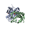
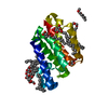
| Structure viewer | Molecule: MolmilJmol/JSmol |
|---|
- Downloads & links
-Download
| PDBx/mmCIF format | 3o1x.cif.gz | 79 KB | Display | PDBx/mmCIF format |
|---|---|---|---|---|
| PDB format | pdb3o1x.ent.gz | 58.5 KB | Display | PDB format |
| PDBx/mmJSON format | 3o1x.json.gz | Tree view | PDBx/mmJSON format | |
| Others |  Other downloads Other downloads |
-Validation report
| Arichive directory | https://data.pdbj.org/pub/pdb/validation_reports/o1/3o1xftp://data.pdbj.org/pub/pdb/validation_reports/o1/3o1x | HTTPS FTP |
|---|
-Related structure data
| Related structure data |  3o1cC  3o1zC  3llj C: citing same article ( S: Starting model for refinement |
|---|---|
| Similar structure data |


-Links
 PDBj
PDBj


- Assembly
| Deposited unit | 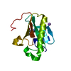
| ||||||||
|---|---|---|---|---|---|---|---|---|---|
| 1 | 
| ||||||||
| Unit cell |
|
-Components
| #1: Protein | Mass: 13681.770 Da / Num. of mol.: 1 / Mutation: C84A Source method: isolated from a genetically manipulated source Source: (gene. exp.)  |
|---|---|
| #2: Chemical | ChemComp-ADN /   Mass: 267.241 Da / Num. of mol.: 1 / Source method: obtained synthetically / Formula: C10H13N5O4 Mass: 267.241 Da / Num. of mol.: 1 / Source method: obtained synthetically / Formula: C10H13N5O4 |
| #3: Chemical | ChemComp-NA /   Mass: 22.990 Da / Num. of mol.: 1 / Source method: obtained synthetically / Formula: Na Mass: 22.990 Da / Num. of mol.: 1 / Source method: obtained synthetically / Formula: Na |
| #4: Water | ChemComp-HOH /  Mass: 18.015 Da / Num. of mol.: 210 / Source method: isolated from a natural source / Formula: H2O Mass: 18.015 Da / Num. of mol.: 210 / Source method: isolated from a natural source / Formula: H2O |
-Experimental details
-Experiment
| Experiment | Method: X-RAY DIFFRACTION / Number of used crystals: 1 |
|---|
- Sample preparation
| Crystal | Density Matthews: 2.04 Å3/Da / Density % sol: 39.79 % |
|---|---|
| Crystal grow | Temperature: 281 K / Method: vapor diffusion, hanging drop / pH: 6.5 Details: 30% PEG 8000, 0.1M sodium cacodylate pH 6.5, O.1M sodium acetate, protein concentration 10 mg/ml, VAPOR DIFFUSION, HANGING DROP, temperature 281K |
-Data collection
| Diffraction | Mean temperature: 100 K |
|---|---|
| Diffraction source | Source: SYNCHROTRON / Site: MAX II  / Beamline: I911-2 / Wavelength: 1.0379 Å / Beamline: I911-2 / Wavelength: 1.0379 Å |
| Detector | Type: MAR CCD 165 mm / Detector: CCD / Date: Nov 19, 2009 / Details: mirrors |
| Radiation | Monochromator: Bent Si (111) crystal, horizontally focusing / Protocol: SINGLE WAVELENGTH / Monochromatic (M) / Laue (L): M / Scattering type: x-ray |
| Radiation wavelength | Wavelength: 1.0379 Å / Relative weight: 1 |
| Reflection | Resolution: 1.08→50 Å / Num. obs: 49845 / % possible obs: 99.6 % / Observed criterion σ(F): 0 / Observed criterion σ(I): -3 / Redundancy: 10.7 % / Biso Wilson estimate: 11 Å2 / Rmerge(I) obs: 0.11 / Net I/σ(I): 16.15 |
| Reflection shell | Resolution: 1.08→1.1 Å / Redundancy: 7.3 % / Rmerge(I) obs: 0.628 / Mean I/σ(I) obs: 2.88 / Num. unique all: 2434 / % possible all: 99.7 |
- Processing
| Software |
| ||||||||||||||||||||||||||||||||||||||||||||||||||||||||||||||||||||||
|---|---|---|---|---|---|---|---|---|---|---|---|---|---|---|---|---|---|---|---|---|---|---|---|---|---|---|---|---|---|---|---|---|---|---|---|---|---|---|---|---|---|---|---|---|---|---|---|---|---|---|---|---|---|---|---|---|---|---|---|---|---|---|---|---|---|---|---|---|---|---|---|
| Refinement | Method to determine structure: MOLECULAR REPLACEMENT Starting model: PDB ENTRY 3LLJ 3llj Resolution: 1.08→23.05 Å / Cor.coef. Fo:Fc: 0.977 / Cor.coef. Fo:Fc free: 0.959 / SU B: 0.815 / SU ML: 0.018 / Cross valid method: THROUGHOUT / σ(F): 0 / ESU R Free: 0.031 / Stereochemistry target values: MAXIMUM LIKELIHOOD / Details: HYDROGENS HAVE BEEN ADDED IN THE RIDING POSITIONS
| ||||||||||||||||||||||||||||||||||||||||||||||||||||||||||||||||||||||
| Solvent computation | Ion probe radii: 0.8 Å / Shrinkage radii: 0.8 Å / VDW probe radii: 1.4 Å / Solvent model: BABINET MODEL WITH MASK | ||||||||||||||||||||||||||||||||||||||||||||||||||||||||||||||||||||||
| Displacement parameters | Biso mean: 23.216 Å2
| ||||||||||||||||||||||||||||||||||||||||||||||||||||||||||||||||||||||
| Refinement step | Cycle: LAST / Resolution: 1.08→23.05 Å
| ||||||||||||||||||||||||||||||||||||||||||||||||||||||||||||||||||||||
| Refine LS restraints |
| ||||||||||||||||||||||||||||||||||||||||||||||||||||||||||||||||||||||
| LS refinement shell | Resolution: 1.08→1.108 Å / Total num. of bins used: 20
|
