 Movie
Movie Controller

[English] 日本語
 Yorodumi
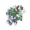
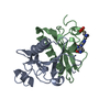
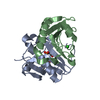
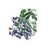
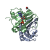
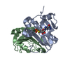
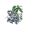
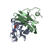
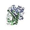
- PDB-1rzy: Crystal structure of rabbit Hint complexed with N-ethylsulfamoyla... -
+ Open data

- Basic information
| Entry | Database: PDB / ID: 1rzy | ||||||
|---|---|---|---|---|---|---|---|
| Title | Crystal structure of rabbit Hint complexed with N-ethylsulfamoyladenosine | ||||||
 Components Components | Histidine triad nucleotide-binding protein 1 | ||||||
 Keywords Keywords | HYDROLASE / HIT protein / protein-inhibitor complex | ||||||
| Function / homology |  Function and homology information Function and homology informationpurine ribonucleotide catabolic process / Hydrolases; Acting on phosphorus-nitrogen bonds / adenosine 5'-monophosphoramidase activity / deSUMOylase activity / protein desumoylation / histone deacetylase complex / intrinsic apoptotic signaling pathway by p53 class mediator / Hydrolases; Acting on peptide bonds (peptidases); Cysteine endopeptidases / nucleotide binding / hydrolase activity ...purine ribonucleotide catabolic process / Hydrolases; Acting on phosphorus-nitrogen bonds / adenosine 5'-monophosphoramidase activity / deSUMOylase activity / protein desumoylation / histone deacetylase complex / intrinsic apoptotic signaling pathway by p53 class mediator / Hydrolases; Acting on peptide bonds (peptidases); Cysteine endopeptidases / nucleotide binding / hydrolase activity / regulation of DNA-templated transcription / proteolysis / nucleus / cytoplasm Similarity search - Function | ||||||
| Biological species |  | ||||||
| Method |  X-RAY DIFFRACTION / FOURIER SYNTHESIS / Resolution: 1.8 Å X-RAY DIFFRACTION / FOURIER SYNTHESIS / Resolution: 1.8 Å | ||||||
 Authors Authors | Krakowiak, A.K. / Pace, H.C. / Blackburn, G.M. / Adams, M. / Mekhalfia, A. / Kaczmarek, R. / Baraniak, J. / Stec, W.J. / Brenner, C. | ||||||
 Citation Citation | Journal: J.Biol.Chem. / Year: 2004 Title: Biochemical, crystallographic, and mutagenic characterization of hint, the AMP-lysine hydrolase, with novel substrates and inhibitors Authors: Krakowiak, A.K. / Pace, H.C. / Blackburn, G.M. / Adams, M. / Mekhalfia, A. / Kaczmarek, R. / Baraniak, J. / Stec, W.J. / Brenner, C. #1: Journal: Nat.Struct.Biol. / Year: 1997Title: Crystal structures of HINT demonstrate that histidine triad proteins are GalT-related nucleotide-binding proteins Authors: Brenner, C. / Garrison, P. / Gilmour, J. / Peisach, D. / Ringe, D. / Petsko, G.A. / Lowenstein, J.M. | ||||||
| History |
|
- Structure visualization
| Structure viewer | Molecule: MolmilJmol/JSmol |
|---|
- Downloads & links
-Download
| PDBx/mmCIF format | 1rzy.cif.gz | 36.7 KB | Display | PDBx/mmCIF format |
|---|---|---|---|---|
| PDB format | pdb1rzy.ent.gz | 24.7 KB | Display | PDB format |
| PDBx/mmJSON format | 1rzy.json.gz | Tree view | PDBx/mmJSON format | |
| Others |  Other downloads Other downloads |
-Validation report
| Arichive directory | https://data.pdbj.org/pub/pdb/validation_reports/rz/1rzyftp://data.pdbj.org/pub/pdb/validation_reports/rz/1rzy | HTTPS FTP |
|---|
-Related structure data
| Related structure data | |
|---|---|
| Similar structure data |


-Links
 PDBj
PDBj


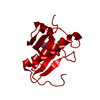
- Assembly
| Deposited unit | 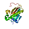
| ||||||||||
|---|---|---|---|---|---|---|---|---|---|---|---|
| 1 | 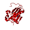
| ||||||||||
| Unit cell |
|
-Components
| #1: Protein | Mass: 13713.835 Da / Num. of mol.: 1 Source method: isolated from a genetically manipulated source Source: (gene. exp.)  |
|---|---|
| #2: Chemical | ChemComp-5AS /   Mass: 374.373 Da / Num. of mol.: 1 / Source method: obtained synthetically / Formula: C12H18N6O6S Mass: 374.373 Da / Num. of mol.: 1 / Source method: obtained synthetically / Formula: C12H18N6O6S |
| #3: Water | ChemComp-HOH /  Mass: 18.015 Da / Num. of mol.: 70 / Source method: isolated from a natural source / Formula: H2O Mass: 18.015 Da / Num. of mol.: 70 / Source method: isolated from a natural source / Formula: H2O |
-Experimental details
-Experiment
| Experiment | Method: X-RAY DIFFRACTION / Number of used crystals: 1 |
|---|
- Sample preparation
| Crystal | Density Matthews: 2.1 Å3/Da / Density % sol: 41.1 % | |||||||||||||||||||||||||
|---|---|---|---|---|---|---|---|---|---|---|---|---|---|---|---|---|---|---|---|---|---|---|---|---|---|---|
| Crystal grow | Temperature: 298 K / Method: vapor diffusion / pH: 7.4 Details: sodium acetate, cacodylate, PEG8000, pH 7.4, VAPOR DIFFUSION, temperature 298K | |||||||||||||||||||||||||
| Crystal grow | *PLUS Method: vapor diffusion / Details: Brenner, C., (1997) Nat.Struct.Biol., 4, 231. | |||||||||||||||||||||||||
| Components of the solutions | *PLUS
|
-Data collection
| Diffraction | Mean temperature: 100 K |
|---|---|
| Diffraction source | Source: ROTATING ANODE / Type: RIGAKU RU200 / Wavelength: 1.5418 Å |
| Detector | Type: RIGAKU RAXIS IV / Detector: IMAGE PLATE / Date: Oct 26, 2002 / Details: osmic confocal blue |
| Radiation | Monochromator: Osmic Confocal mirrors / Protocol: SINGLE WAVELENGTH / Monochromatic (M) / Laue (L): M / Scattering type: x-ray |
| Radiation wavelength | Wavelength: 1.5418 Å / Relative weight: 1 |
| Reflection | Resolution: 1.8→23 Å / Num. obs: 10698 / % possible obs: 93.9 % / Observed criterion σ(F): 2 / Observed criterion σ(I): 1 / Redundancy: 8.4 % / Biso Wilson estimate: 16.9 Å2 / Rsym value: 0.0065 / Net I/σ(I): 9.2 |
| Reflection shell | Resolution: 1.8→1.86 Å / Redundancy: 3.4 % / Mean I/σ(I) obs: 5.1 / Num. unique all: 968 / Rsym value: 0.143 / % possible all: 88.3 |
| Reflection | *PLUS Highest resolution: 1.8 Å / Lowest resolution: 23 Å / Num. measured all: 95355 / Rmerge(I) obs: 0.065 |
| Reflection shell | *PLUS % possible obs: 88.3 % / Rmerge(I) obs: 0.143 |
- Processing
| Software |
| ||||||||||||||||||||||||||||||||||||
|---|---|---|---|---|---|---|---|---|---|---|---|---|---|---|---|---|---|---|---|---|---|---|---|---|---|---|---|---|---|---|---|---|---|---|---|---|---|
| Refinement | Method to determine structure: FOURIER SYNTHESIS / Resolution: 1.8→23 Å / Cross valid method: THROUGHOUT / σ(F): 0 / Stereochemistry target values: Engh & Huber
| ||||||||||||||||||||||||||||||||||||
| Refinement step | Cycle: LAST / Resolution: 1.8→23 Å
| ||||||||||||||||||||||||||||||||||||
| Refine LS restraints |
| ||||||||||||||||||||||||||||||||||||
| Refinement | *PLUS Highest resolution: 1.8 Å / Lowest resolution: 23 Å / % reflection Rfree: 7 % | ||||||||||||||||||||||||||||||||||||
| Solvent computation | *PLUS | ||||||||||||||||||||||||||||||||||||
| Displacement parameters | *PLUS | ||||||||||||||||||||||||||||||||||||
| Refine LS restraints | *PLUS
| ||||||||||||||||||||||||||||||||||||
| LS refinement shell | *PLUS Rfactor Rfree: 0.281 / Rfactor Rwork: 0.299 |
