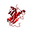
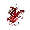
Mass: 12584.530 Da / Num. of mol.: 1 Source method: isolated from a genetically manipulated source Source: (gene. exp.) Oryctolagus cuniculus (rabbit) Description: RABBIT HINT CDNA WAS CLONED FROM HEART LIBRARY, EXPRESSED IN ESCHERICHIA COLI, AND PURIFIED BY ADENOSINE-AGAROSE AFFINITY CHROMATOGRAPHY Gene: HINT / Organ: HEART / Plasmid: PSGA02-HINT / Gene (production host): HINT / Production host: Escherichia coli (E. coli) / Strain (production host): JM109/DE3/LACIQ / References: UniProt: P80912
Mass: 18.015 Da / Num. of mol.: 51 / Source method: isolated from a natural source / Formula: H2O
-
Experimental details
-
Experiment
Experiment
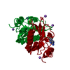
Method: X-RAY DIFFRACTION / Number of used crystals: 1
-
Sample preparation
Crystal
Density Matthews: 2.08 Å3/Da / Density % sol: 41 % Description: 5RHN (TETRAGONAL HINT-8-BR-AMP, ISOMORPHOUS WITH HINT WITHOUT NUCLEOTIDE) WAS DETERMINED BY REFINEMENT OF HINT WITHOUT NUCLEOTIDE.
Crystal grow
pH: 6.5 Details: 30% POLYETHYLENE GLYCOL 8000, 0.1 M SODIUM ACETATE, 0.1 M SODIUM CACODYLATE, PH 6.5
In the structure databanks used in Yorodumi, some data are registered as the other names, "COVID-19 virus" and "2019-nCoV". Here are the details of the virus and the list of structure data.
Jan 31, 2019. EMDB accession codes are about to change! (news from PDBe EMDB page)
EMDB accession codes are about to change! (news from PDBe EMDB page)
The allocation of 4 digits for EMDB accession codes will soon come to an end. Whilst these codes will remain in use, new EMDB accession codes will include an additional digit and will expand incrementally as the available range of codes is exhausted. The current 4-digit format prefixed with “EMD-” (i.e. EMD-XXXX) will advance to a 5-digit format (i.e. EMD-XXXXX), and so on. It is currently estimated that the 4-digit codes will be depleted around Spring 2019, at which point the 5-digit format will come into force.
The EM Navigator/Yorodumi systems omit the EMD- prefix.
Related info.:Q: What is EMD? / ID/Accession-code notation in Yorodumi/EM Navigator
Yorodumi is a browser for structure data from EMDB, PDB, SASBDB, etc.
This page is also the successor to EM Navigator detail page, and also detail information page/front-end page for Omokage search.
The word "yorodu" (or yorozu) is an old Japanese word meaning "ten thousand". "mi" (miru) is to see.
Related info.:EMDB / PDB / SASBDB / Comparison of 3 databanks / Yorodumi Search / Aug 31, 2016. New EM Navigator & Yorodumi / Yorodumi Papers / Jmol/JSmol / Function and homology information / Changes in new EM Navigator and Yorodumi
 Movie
Movie Controller

 Yorodumi
 Open data
 Basic information
 Components
 Keywords
 Function and homology information
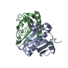
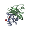
 X-RAY DIFFRACTION / Resolution: 2.31 Å
  Authors
 Citation
 Structure visualization
 Downloads & links
 Other downloads


 PDBj
PDBj


 Assembly


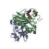
 Mass: 426.117 Da / Num. of mol.: 1 / Source method: obtained synthetically / Formula: C10H13BrN5O7P
 Mass: 18.015 Da / Num. of mol.: 51 / Source method: isolated from a natural source / Formula: H2O
 Sample preparation
 Processing