 Movie
Movie Controller


+ Open data

- Basic information
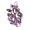
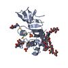
| Entry | Database: PDB / ID: 4myv | ||||||
|---|---|---|---|---|---|---|---|
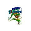
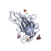
| Title | Free HSV-2 gD structure | ||||||
 Components Components | Envelope glycoprotein D | ||||||
 Keywords Keywords | VIRAL PROTEIN / IgV-like core / N-/C-terminal extensions / receptor binding / nectin-1 / HVEM / viral surface | ||||||
| Function / homology |  Function and homology information Function and homology informationcoreceptor-mediated virion attachment to host cell / entry receptor-mediated virion attachment to host cell / receptor ligand activity / fusion of virus membrane with host plasma membrane / viral envelope / symbiont entry into host cell / virion membrane / membrane / metal ion binding Similarity search - Function | ||||||
| Biological species |   Human herpesvirus 2 Human herpesvirus 2 | ||||||
| Method |  X-RAY DIFFRACTION / SYNCHROTRON / MOLECULAR REPLACEMENT / Resolution: 1.801 Å X-RAY DIFFRACTION / SYNCHROTRON / MOLECULAR REPLACEMENT / Resolution: 1.801 Å | ||||||
 Authors Authors | Lu, G. / Zhang, N. / Qi, J. / Li, Y. / Chen, Z. / Zheng, C. / Yan, J. / Gao, G.F. | ||||||
 Citation Citation | Journal: J.Virol. / Year: 2014 Title: Crystal structure of herpes simplex virus 2 gD bound to nectin-1 reveals a conserved mode of receptor recognition. Authors: Lu, G. / Zhang, N. / Qi, J. / Li, Y. / Chen, Z. / Zheng, C. / Gao, G.F. / Yan, J. | ||||||
| History |
|
- Structure visualization
| Structure viewer | Molecule: MolmilJmol/JSmol |
|---|
- Downloads & links
-Download
| PDBx/mmCIF format | 4myv.cif.gz | 97.8 KB | Display | PDBx/mmCIF format |
|---|---|---|---|---|
| PDB format | pdb4myv.ent.gz | 72 KB | Display | PDB format |
| PDBx/mmJSON format | 4myv.json.gz | Tree view | PDBx/mmJSON format | |
| Others |  Other downloads Other downloads |
-Validation report
| Arichive directory | https://data.pdbj.org/pub/pdb/validation_reports/my/4myvftp://data.pdbj.org/pub/pdb/validation_reports/my/4myv | HTTPS FTP |
|---|
-Related structure data
| Related structure data |  4mywC  1l2gS C: citing same article ( S: Starting model for refinement |
|---|---|
| Similar structure data |


-Links
 PDBj
PDBj- Assembly
| Deposited unit | 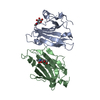
| ||||||||
|---|---|---|---|---|---|---|---|---|---|
| 1 | 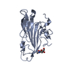
| ||||||||
| 2 | 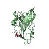
| ||||||||
| Unit cell |
|
-Components
| #1: Protein | Mass: 32994.465 Da / Num. of mol.: 2 / Fragment: UNP residues 26-310 Source method: isolated from a genetically manipulated source Source: (gene. exp.) Human herpesvirus 2 / Strain: 333 / Gene: gD, US6 / Plasmid: pFast-bac1 / Cell (production host): High5 / Cell line (production host): BTI-TN-5B1-4 / Production host:  Trichoplusia ni (cabbage looper) / References: UniProt: P03172 Trichoplusia ni (cabbage looper) / References: UniProt: P03172#2: Sugar |   Type: D-saccharide, beta linking / Mass: 221.208 Da / Num. of mol.: 2 Type: D-saccharide, beta linking / Mass: 221.208 Da / Num. of mol.: 2Source method: isolated from a genetically manipulated source Formula: C8H15NO6 #3: Water | ChemComp-HOH / |  Mass: 18.015 Da / Num. of mol.: 407 / Source method: isolated from a natural source / Formula: H2O Mass: 18.015 Da / Num. of mol.: 407 / Source method: isolated from a natural source / Formula: H2OHas protein modification | Y | Sequence details | NATURAL VARIANT AT THIS POSITION. THE SEQUENCE IS BASED ON A RECORD IN GENBANK WITH THE ACCESSION ...NATURAL VARIANT AT THIS POSITION. THE SEQUENCE IS BASED ON A RECORD IN GENBANK WITH THE ACCESSION NUMBER EU018091 | |
|---|
-Experimental details
-Experiment
| Experiment | Method: X-RAY DIFFRACTION / Number of used crystals: 1 |
|---|
- Sample preparation
| Crystal grow | Temperature: 291 K / Method: vapor diffusion, hanging drop / pH: 7.2 Details: 0.1mol/L Hepes pH 7.2, 5% (V/V) MPD, 10% PEG 10000, VAPOR DIFFUSION, HANGING DROP, temperature 291K |
|---|
-Data collection
| Diffraction | Mean temperature: 100 K |
|---|---|
| Diffraction source | Source: SYNCHROTRON / Site: SSRF  / Beamline: BL17U / Wavelength: 0.97916 Å / Beamline: BL17U / Wavelength: 0.97916 Å |
| Detector | Type: ADSC QUANTUM 315r / Detector: CCD / Date: Nov 8, 2010 |
| Radiation | Monochromator: GRAPHITE / Protocol: SINGLE WAVELENGTH / Monochromatic (M) / Laue (L): M / Scattering type: x-ray |
| Radiation wavelength | Wavelength: 0.97916 Å / Relative weight: 1 |
| Reflection | Resolution: 1.8→50 Å / Num. all: 41946 / Num. obs: 40614 / % possible obs: 96.8 % / Observed criterion σ(F): 0 / Observed criterion σ(I): -3 / Redundancy: 3.9 % / Rmerge(I) obs: 0.085 / Rsym value: 0.085 / Net I/σ(I): 15.89 |
| Reflection shell | Resolution: 1.8→1.86 Å / Redundancy: 2.8 % / Rmerge(I) obs: 0.297 / Mean I/σ(I) obs: 2.714 / Num. unique all: 3384 / Rsym value: 0.297 / % possible all: 81.2 |
- Processing
| Software |
| ||||||||||||||||||||||||||||||||||||||||||||||||||||||||||||||||||||||||||||||||||||||||||||||||
|---|---|---|---|---|---|---|---|---|---|---|---|---|---|---|---|---|---|---|---|---|---|---|---|---|---|---|---|---|---|---|---|---|---|---|---|---|---|---|---|---|---|---|---|---|---|---|---|---|---|---|---|---|---|---|---|---|---|---|---|---|---|---|---|---|---|---|---|---|---|---|---|---|---|---|---|---|---|---|---|---|---|---|---|---|---|---|---|---|---|---|---|---|---|---|---|---|---|
| Refinement | Method to determine structure: MOLECULAR REPLACEMENT Starting model: 1L2G Resolution: 1.801→36.268 Å / SU ML: 0.19 / σ(F): 1.34 / Phase error: 21.48 / Stereochemistry target values: Engh & Huber
| ||||||||||||||||||||||||||||||||||||||||||||||||||||||||||||||||||||||||||||||||||||||||||||||||
| Solvent computation | Shrinkage radii: 0.9 Å / VDW probe radii: 1.11 Å / Solvent model: FLAT BULK SOLVENT MODEL | ||||||||||||||||||||||||||||||||||||||||||||||||||||||||||||||||||||||||||||||||||||||||||||||||
| Refinement step | Cycle: LAST / Resolution: 1.801→36.268 Å
| ||||||||||||||||||||||||||||||||||||||||||||||||||||||||||||||||||||||||||||||||||||||||||||||||
| Refine LS restraints |
| ||||||||||||||||||||||||||||||||||||||||||||||||||||||||||||||||||||||||||||||||||||||||||||||||
| LS refinement shell | Refine-ID: X-RAY DIFFRACTION / Total num. of bins used: 15
|
