 Movie
Movie Controller

[English] 日本語
 Yorodumi
- PDB-3u82: Binding of herpes simplex virus glycoprotein D to nectin-1 exploi... -
+ Open data

- Basic information
| Entry | Database: PDB / ID: 3u82 | ||||||
|---|---|---|---|---|---|---|---|
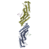
| Title | Binding of herpes simplex virus glycoprotein D to nectin-1 exploits host cell adhesion | ||||||
 Components Components |
| ||||||
 Keywords Keywords | VIRAL PROTEIN/CELL ADHESION / HSV-1 gD / nectin-1 / binding mode / nectin-1 dimerization preclusion / VIRAL PROTEIN-CELL ADHESION complex | ||||||
| Function / homology |  Function and homology information Function and homology informationdesmosome organization / Nectin/Necl trans heterodimerization / protein localization to cell junction / lens morphogenesis in camera-type eye / enamel mineralization / growth cone membrane / cell adhesion mediator activity / cochlea morphogenesis / cell-cell contact zone / virion binding ...desmosome organization / Nectin/Necl trans heterodimerization / protein localization to cell junction / lens morphogenesis in camera-type eye / enamel mineralization / growth cone membrane / cell adhesion mediator activity / cochlea morphogenesis / cell-cell contact zone / virion binding / Adherens junctions interactions / heterophilic cell-cell adhesion / apical junction complex / regulation of synapse assembly / homophilic cell-cell adhesion / coreceptor activity / presynaptic active zone membrane / cell adhesion molecule binding / axon guidance / hippocampal mossy fiber to CA3 synapse / adherens junction / iron ion transport / cell-cell adhesion / carbohydrate binding / virus receptor activity / retina development in camera-type eye / host cell Golgi apparatus / entry receptor-mediated virion attachment to host cell / cell adhesion / immune response / receptor ligand activity / viral envelope / symbiont entry into host cell / dendrite / protein-containing complex binding / virion membrane / protein homodimerization activity / extracellular region / membrane / metal ion binding / identical protein binding / plasma membrane Similarity search - Function | ||||||
| Biological species |   Human herpesvirus 1 (Herpes simplex virus type 1) Human herpesvirus 1 (Herpes simplex virus type 1) Homo sapiens (human) Homo sapiens (human) | ||||||
| Method |  X-RAY DIFFRACTION / SYNCHROTRON / MOLECULAR REPLACEMENT / Resolution: 3.164 Å X-RAY DIFFRACTION / SYNCHROTRON / MOLECULAR REPLACEMENT / Resolution: 3.164 Å | ||||||
 Authors Authors | Zhang, N. / Yan, J. / Lu, G. / Guo, Z. / Fan, Z. / Wang, J. / Shi, Y. / Qi, J. / Gao, G.F. | ||||||
 Citation Citation | Journal: Nat Commun / Year: 2011 Title: Binding of herpes simplex virus glycoprotein D to nectin-1 exploits host cell adhesion. Authors: Zhang, N. / Yan, J. / Lu, G. / Guo, Z. / Fan, Z. / Wang, J. / Shi, Y. / Qi, J. / Gao, G.F. | ||||||
| History |
|
- Structure visualization
| Structure viewer | Molecule: MolmilJmol/JSmol |
|---|
- Downloads & links
-Download
| PDBx/mmCIF format | 3u82.cif.gz | 228.4 KB | Display | PDBx/mmCIF format |
|---|---|---|---|---|
| PDB format | pdb3u82.ent.gz | 186.5 KB | Display | PDB format |
| PDBx/mmJSON format | 3u82.json.gz | Tree view | PDBx/mmJSON format | |
| Others |  Other downloads Other downloads |
-Validation report
| Arichive directory | https://data.pdbj.org/pub/pdb/validation_reports/u8/3u82ftp://data.pdbj.org/pub/pdb/validation_reports/u8/3u82 | HTTPS FTP |
|---|
-Related structure data
| Related structure data |  3u83C 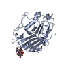 2c36S  3alpS S: Starting model for refinement C: citing same article ( |
|---|---|
| Similar structure data |
-Links
 PDBj
PDBj


- Assembly
| Deposited unit | 
| ||||||||
|---|---|---|---|---|---|---|---|---|---|
| 1 |
| ||||||||
| Unit cell |
|
-Components
| #1: Protein | Mass: 32481.893 Da / Num. of mol.: 1 / Fragment: UNP RESIDUES 26-310 Source method: isolated from a genetically manipulated source Source: (gene. exp.) Human herpesvirus 1 (Herpes simplex virus type 1)Strain: 17 / Gene: gD, US6 / Production host:   Spodoptera frugiperda (fall armyworm) / References: UniProt: Q69091 Spodoptera frugiperda (fall armyworm) / References: UniProt: Q69091 |
|---|---|
| #2: Protein | Mass: 35488.844 Da / Num. of mol.: 1 / Fragment: UNP RESIDUES 30-335 Source method: isolated from a genetically manipulated source Source: (gene. exp.) Homo sapiens (human) / Gene: PVRL1, HVEC, PRR1 / Production host:  |
| Has protein modification | Y |
-Experimental details
-Experiment
| Experiment | Method: X-RAY DIFFRACTION / Number of used crystals: 1 |
|---|
- Sample preparation
| Crystal | Density Matthews: 3.07 Å3/Da / Density % sol: 59.92 % |
|---|---|
| Crystal grow | Temperature: 277 K / Method: vapor diffusion, hanging drop / pH: 5.5 Details: 20% PEG 1000, 0.1M lithium sulfate monohydrate, 0.1M sodium citrate tribasic dehydrate, pH 5.5 , VAPOR DIFFUSION, HANGING DROP, temperature 277K |
-Data collection
| Diffraction | Mean temperature: 100 K |
|---|---|
| Diffraction source | Source: SYNCHROTRON / Site: SSRF  / Beamline: BL17U / Wavelength: 0.97916 Å / Beamline: BL17U / Wavelength: 0.97916 Å |
| Detector | Type: ADSC QUANTUM 315 / Detector: CCD / Date: Jul 19, 2011 |
| Radiation | Monochromator: GRAPHITE / Protocol: SINGLE WAVELENGTH / Monochromatic (M) / Laue (L): M / Scattering type: x-ray |
| Radiation wavelength | Wavelength: 0.97916 Å / Relative weight: 1 |
| Reflection | Resolution: 3.164→50 Å / Num. all: 13572 / Num. obs: 13572 / % possible obs: 94.9 % / Observed criterion σ(F): 2 / Observed criterion σ(I): 2 / Redundancy: 4.2 % / Rmerge(I) obs: 0.102 / Net I/σ(I): 14.4 |
| Reflection shell | Resolution: 3.2→3.31 Å / Redundancy: 3.9 % / Rmerge(I) obs: 0.504 / Mean I/σ(I) obs: 1.7 / Num. unique all: 1158 / % possible all: 82.7 |
- Processing
| Software |
| ||||||||||||||||||||||||||||||
|---|---|---|---|---|---|---|---|---|---|---|---|---|---|---|---|---|---|---|---|---|---|---|---|---|---|---|---|---|---|---|---|
| Refinement | Method to determine structure: MOLECULAR REPLACEMENT Starting model: 2C36, 3ALP Resolution: 3.164→38.378 Å / SU ML: 0.5 / σ(F): 0.04 / Phase error: 43.12 / Stereochemistry target values: ML
| ||||||||||||||||||||||||||||||
| Solvent computation | Shrinkage radii: 0.9 Å / VDW probe radii: 1.11 Å / Solvent model: FLAT BULK SOLVENT MODEL / Bsol: 130.701 Å2 / ksol: 0.304 e/Å3 | ||||||||||||||||||||||||||||||
| Displacement parameters |
| ||||||||||||||||||||||||||||||
| Refinement step | Cycle: LAST / Resolution: 3.164→38.378 Å
| ||||||||||||||||||||||||||||||
| Refine LS restraints |
| ||||||||||||||||||||||||||||||
| LS refinement shell | Refine-ID: X-RAY DIFFRACTION / Total num. of bins used: 4
|
