 Movie
Movie Controller


+ Open data

- Basic information
| Entry | Database: PDB / ID: 4myw | |||||||||
|---|---|---|---|---|---|---|---|---|---|---|
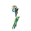
| Title | Structure of HSV-2 gD bound to nectin-1 | |||||||||
 Components Components |
| |||||||||
 Keywords Keywords | VIRAL PROTEIN/CELL ADHESION / IgV-like core / N-/C-terminal extensions / tandem arranged V-C-C Ig-like domains / receptor binding / cell surface attachment / viral / cellular surface / VIRAL PROTEIN-CELL ADHESION complex | |||||||||
| Function / homology |  Function and homology information Function and homology informationcoreceptor-mediated virion attachment to host cell / desmosome organization / Nectin/Necl trans heterodimerization / protein localization to cell junction / lens morphogenesis in camera-type eye / enamel mineralization / growth cone membrane / cell adhesion mediator activity / cochlea morphogenesis / cell-cell contact zone ...coreceptor-mediated virion attachment to host cell / desmosome organization / Nectin/Necl trans heterodimerization / protein localization to cell junction / lens morphogenesis in camera-type eye / enamel mineralization / growth cone membrane / cell adhesion mediator activity / cochlea morphogenesis / cell-cell contact zone / virion binding / Adherens junctions interactions / heterophilic cell-cell adhesion / apical junction complex / regulation of synapse assembly / homophilic cell-cell adhesion / coreceptor activity / presynaptic active zone membrane / cell adhesion molecule binding / axon guidance / hippocampal mossy fiber to CA3 synapse / adherens junction / iron ion transport / cell-cell adhesion / carbohydrate binding / virus receptor activity / retina development in camera-type eye / entry receptor-mediated virion attachment to host cell / cell adhesion / immune response / receptor ligand activity / fusion of virus membrane with host plasma membrane / viral envelope / symbiont entry into host cell / dendrite / protein-containing complex binding / virion membrane / protein homodimerization activity / extracellular region / membrane / metal ion binding / identical protein binding / plasma membrane Similarity search - Function | |||||||||
| Biological species |  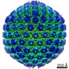 Human herpesvirus 2 Human herpesvirus 2 Homo sapiens (human) Homo sapiens (human) | |||||||||
| Method |  X-RAY DIFFRACTION / SYNCHROTRON / MOLECULAR REPLACEMENT / Resolution: 3.189 Å X-RAY DIFFRACTION / SYNCHROTRON / MOLECULAR REPLACEMENT / Resolution: 3.189 Å | |||||||||
 Authors Authors | Lu, G. / Zhang, N. / Qi, J. / Li, Y. / Chen, Z. / Zheng, C. / Yan, J. / Gao, G.F. | |||||||||
 Citation Citation | Journal: J.Virol. / Year: 2014 Title: Crystal structure of herpes simplex virus 2 gD bound to nectin-1 reveals a conserved mode of receptor recognition. Authors: Lu, G. / Zhang, N. / Qi, J. / Li, Y. / Chen, Z. / Zheng, C. / Gao, G.F. / Yan, J. | |||||||||
| History |
|
- Structure visualization
| Structure viewer | Molecule: MolmilJmol/JSmol |
|---|
- Downloads & links
-Download
| PDBx/mmCIF format | 4myw.cif.gz | 436.6 KB | Display | PDBx/mmCIF format |
|---|---|---|---|---|
| PDB format | pdb4myw.ent.gz | 359.1 KB | Display | PDB format |
| PDBx/mmJSON format | 4myw.json.gz | Tree view | PDBx/mmJSON format | |
| Others |  Other downloads Other downloads |
-Validation report
| Arichive directory | https://data.pdbj.org/pub/pdb/validation_reports/my/4mywftp://data.pdbj.org/pub/pdb/validation_reports/my/4myw | HTTPS FTP |
|---|
-Related structure data
| Related structure data | 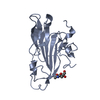 4myvC 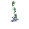 3u82S S: Starting model for refinement C: citing same article ( |
|---|---|
| Similar structure data |


-Links
 PDBj
PDBj


- Assembly
| Deposited unit | 
| ||||||||
|---|---|---|---|---|---|---|---|---|---|
| 1 | 
| ||||||||
| 2 | 
| ||||||||
| Unit cell |
|
-Components
| #1: Protein | Mass: 32994.465 Da / Num. of mol.: 2 / Fragment: UNP RESIDUES 26-310 Source method: isolated from a genetically manipulated source Source: (gene. exp.) Human herpesvirus 2 / Strain: 333 / Gene: gD, US6 / Plasmid: pFast-bac1 / Cell (production host): High5 / Cell line (production host): BTI-TN-5B1-4 / Production host:  Trichoplusia ni (cabbage looper) / References: UniProt: P03172 Trichoplusia ni (cabbage looper) / References: UniProt: P03172#2: Protein | Mass: 36938.492 Da / Num. of mol.: 2 / Fragment: UNP RESIDUES 30-335 Source method: isolated from a genetically manipulated source Source: (gene. exp.) Homo sapiens (human) / Gene: HVEC, nectin-1, PRR1, PVRL1 / Plasmid: pET-21b / Production host:  #3: Polysaccharide | Source method: isolated from a genetically manipulated source #4: Sugar |   Type: D-saccharide, beta linking / Mass: 221.208 Da / Num. of mol.: 2 Type: D-saccharide, beta linking / Mass: 221.208 Da / Num. of mol.: 2Source method: isolated from a genetically manipulated source Formula: C8H15NO6 #5: Water | ChemComp-HOH / |  Mass: 18.015 Da / Num. of mol.: 57 / Source method: isolated from a natural source / Formula: H2O Mass: 18.015 Da / Num. of mol.: 57 / Source method: isolated from a natural source / Formula: H2OHas protein modification | Y | Sequence details | NATURAL VARIANT AT THIS POSITION. THE SEQUENCE IS BASED ON A RECORD IN GENBANK WITH THE ACCESSION ...NATURAL VARIANT AT THIS POSITION. THE SEQUENCE IS BASED ON A RECORD IN GENBANK WITH THE ACCESSION NUMBER EU018091 | |
|---|
-Experimental details
-Experiment
| Experiment | Method: X-RAY DIFFRACTION / Number of used crystals: 1 |
|---|
- Sample preparation
| Crystal | Density Matthews: 3.13 Å3/Da / Density % sol: 60.72 % |
|---|---|
| Crystal grow | Temperature: 291 K / Method: vapor diffusion, sitting drop / pH: 6 Details: 0.2M sodium chloride, 0.1M sodium cacodylate, 8% w/v PEG 8000, pH 6.0, VAPOR DIFFUSION, SITTING DROP, temperature 291K |
-Data collection
| Diffraction | Mean temperature: 100 K |
|---|---|
| Diffraction source | Source: SYNCHROTRON / Site: SSRF  / Beamline: BL17U / Wavelength: 0.97939 Å / Beamline: BL17U / Wavelength: 0.97939 Å |
| Detector | Type: ADSC QUANTUM 315r / Detector: CCD / Date: Apr 12, 2012 |
| Radiation | Monochromator: GRAPHITE / Protocol: SINGLE WAVELENGTH / Monochromatic (M) / Laue (L): M / Scattering type: x-ray |
| Radiation wavelength | Wavelength: 0.97939 Å / Relative weight: 1 |
| Reflection | Resolution: 3.189→50 Å / Num. all: 30221 / Num. obs: 30129 / % possible obs: 99.7 % / Observed criterion σ(F): 0 / Observed criterion σ(I): -3 / Redundancy: 4.1 % / Rmerge(I) obs: 0.091 / Rsym value: 0.091 / Net I/σ(I): 15.672 |
| Reflection shell | Resolution: 3.2→3.31 Å / Redundancy: 3.8 % / Rmerge(I) obs: 0.492 / Mean I/σ(I) obs: 2.884 / Num. unique all: 2969 / Rsym value: 0.492 / % possible all: 99.5 |
- Processing
| Software |
| ||||||||||||||||||||||||||||||||||||||||||||||||||||||||||||||||||
|---|---|---|---|---|---|---|---|---|---|---|---|---|---|---|---|---|---|---|---|---|---|---|---|---|---|---|---|---|---|---|---|---|---|---|---|---|---|---|---|---|---|---|---|---|---|---|---|---|---|---|---|---|---|---|---|---|---|---|---|---|---|---|---|---|---|---|---|
| Refinement | Method to determine structure: MOLECULAR REPLACEMENT Starting model: 3U82 Resolution: 3.189→38.93 Å / SU ML: 0.29 / σ(F): 0.13 / Phase error: 24.55 / Stereochemistry target values: Engh & Huber
| ||||||||||||||||||||||||||||||||||||||||||||||||||||||||||||||||||
| Solvent computation | Shrinkage radii: 0.9 Å / VDW probe radii: 1.11 Å / Solvent model: FLAT BULK SOLVENT MODEL / Bsol: 28.088 Å2 / ksol: 0.294 e/Å3 | ||||||||||||||||||||||||||||||||||||||||||||||||||||||||||||||||||
| Displacement parameters |
| ||||||||||||||||||||||||||||||||||||||||||||||||||||||||||||||||||
| Refinement step | Cycle: LAST / Resolution: 3.189→38.93 Å
| ||||||||||||||||||||||||||||||||||||||||||||||||||||||||||||||||||
| Refine LS restraints |
| ||||||||||||||||||||||||||||||||||||||||||||||||||||||||||||||||||
| LS refinement shell | Refine-ID: X-RAY DIFFRACTION / Total num. of bins used: 10
| ||||||||||||||||||||||||||||||||||||||||||||||||||||||||||||||||||
| Refinement TLS params. | Method: refined / Origin x: -30.84 Å / Origin y: 41.4651 Å / Origin z: -7.6333 Å
| ||||||||||||||||||||||||||||||||||||||||||||||||||||||||||||||||||
| Refinement TLS group | Selection details: ALL |
