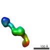
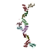
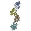
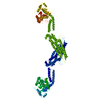
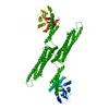
THE BIOLOGICALLY RELEVANT ASSEMBLY IS LINEAR OLIGOMER CREATED BY CRYSTALLOGRAPHIC SYMMETRY ALONG THE C-AXIS OF THE CRYSTAL.THERE ARE TWO INTERFACES INVOLVED IN THE BUILDING OF THE OLIGOMER WHICH IS SEEN IN THE ASSEMBLIES GIVEN IN REMARK 350.
-
Components
#1: Protein
Interferon-inducedGTP-bindingproteinMx1 / Interferon-induced protein p78 / IFI-78K / Interferon-regulated resistance GTP-binding protein MxA ...Interferon-induced protein p78 / IFI-78K / Interferon-regulated resistance GTP-binding protein MxA / Myxoma resistance protein 1 / Myxovirus resistance protein 1 / Interferon-induced GTP-binding protein Mx1 / N-terminally processed
Mass: 69176.812 Da / Num. of mol.: 1 / Fragment: UNP residues 33-662 / Mutation: Y440A,R441A,G442A,R443A Source method: isolated from a genetically manipulated source Source: (gene. exp.) Homo sapiens (human) / Gene: MX1 / Plasmid: pSKB-LNB / Production host: Escherichia coli (E. coli) / Strain (production host): BL21 DE3 Rosetta / References: UniProt: P20591
-
Experimental details
-
Experiment
Experiment
Method: X-RAY DIFFRACTION / Number of used crystals: 1
-
Sample preparation
Crystal
Density Matthews: 4.24 Å3/Da / Density % sol: 70.97 %
Crystal grow
Temperature: 293 K / Method: vapor diffusion, hanging drop / pH: 7.6 Details: 7% PEG3350, 100 mM HEPES (pH 7.6), 80 mM NaCl, 2.5% 2-methyl-2,4-pentandiol (MPD), 5% glycerol, VAPOR DIFFUSION, HANGING DROP, temperature 293K
Resolution: 3.5→34.98 Å / Cor.coef. Fo:Fc: 0.899 / Cor.coef. Fo:Fc free: 0.838 / SU B: 85.978 / SU ML: 0.584 / Cross valid method: THROUGHOUT / σ(F): -3 / ESU R Free: 0.799 / Stereochemistry target values: MAXIMUM LIKELIHOOD / Details: HYDROGENS HAVE BEEN USED IF PRESENT IN THE INPUT
Rfactor
Num. reflection
% reflection
Selection details
Rfree
0.2954
520
5 %
RANDOM
Rwork
0.2616
-
-
-
all
0.26324
14578
-
-
obs
0.26324
9831
71 %
-
Solvent computation
Ion probe radii: 0.8 Å / Shrinkage radii: 0.8 Å / VDW probe radii: 1.2 Å / Solvent model: MASK
Displacement parameters
Biso mean: 140.837 Å2
Baniso -1
Baniso -2
Baniso -3
1-
-2.29 Å2
0 Å2
-1.4 Å2
2-
-
-0.49 Å2
0 Å2
3-
-
-
1.99 Å2
Refinement step
Cycle: LAST / Resolution: 3.5→34.98 Å
Protein
Nucleic acid
Ligand
Solvent
Total
Num. atoms
4507
0
0
0
4507
Refine LS restraints
Refine-ID
Type
Dev ideal
Dev ideal target
Number
X-RAY DIFFRACTION
r_bond_refined_d
0.015
0.022
4572
X-RAY DIFFRACTION
r_bond_other_d
0.003
0.02
3184
X-RAY DIFFRACTION
r_angle_refined_deg
1.583
1.972
6148
X-RAY DIFFRACTION
r_angle_other_deg
1.177
3
7800
X-RAY DIFFRACTION
r_dihedral_angle_1_deg
5.506
5
552
X-RAY DIFFRACTION
r_dihedral_angle_2_deg
34.341
25.067
223
X-RAY DIFFRACTION
r_dihedral_angle_3_deg
14.244
15
897
X-RAY DIFFRACTION
r_dihedral_angle_4_deg
13.978
15
29
X-RAY DIFFRACTION
r_chiral_restr
0.078
0.2
701
X-RAY DIFFRACTION
r_gen_planes_refined
0.007
0.02
4968
X-RAY DIFFRACTION
r_gen_planes_other
0.003
0.02
858
LS refinement shell
Resolution: 3.502→3.592 Å / Total num. of bins used: 20
Rfactor
Num. reflection
% reflection
Rfree
0.326
5
-
Rwork
0.501
51
-
obs
-
-
5.23 %
Refinement TLS params.
Method: refined / Refine-ID: X-RAY DIFFRACTION
ID
L11 (°2)
L12 (°2)
L13 (°2)
L22 (°2)
L23 (°2)
L33 (°2)
S11 (Å °)
S12 (Å °)
S13 (Å °)
S21 (Å °)
S22 (Å °)
S23 (Å °)
S31 (Å °)
S32 (Å °)
S33 (Å °)
T11 (Å2)
T12 (Å2)
T13 (Å2)
T22 (Å2)
T23 (Å2)
T33 (Å2)
Origin x (Å)
Origin y (Å)
Origin z (Å)
1
4.6063
-0.9485
1.5611
5.6096
1.5511
8.719
-0.3805
-0.4068
-1.0368
0.2331
0.3862
-0.5798
0.9185
0.4325
-0.0057
1.0528
0.951
0.0846
1.1055
0.2622
0.67
38.918
-21.474
30.724
2
6.6127
-1.7997
-0.7008
7.8728
-4.724
27.019
-0.3022
-0.3413
-0.5039
0.2429
0.5575
0.3307
0.9152
1.1491
-0.2553
0.5518
0.6168
0.0184
0.8321
-0.0937
0.1578
23.213
-3.981
12.535
3
3.9265
1.4794
-2.8631
6.2095
-7.4698
14.4673
-0.2304
-0.4863
0.5749
-0.1445
0.3086
-0.081
0.2891
0.753
-0.0782
0.248
0.0331
0.0646
0.3448
-0.2197
0.3626
24.484
12.613
-45.562
Refinement TLS group
ID
Refine-ID
Refine TLS-ID
Auth asym-ID
Auth seq-ID
1
X-RAY DIFFRACTION
1
A
64 - 340
2
X-RAY DIFFRACTION
2
A
1 - 63
3
X-RAY DIFFRACTION
2
A
341 - 365
4
X-RAY DIFFRACTION
2
A
638 - 662
5
X-RAY DIFFRACTION
3
A
366 - 637
+
About Yorodumi
-
News
-
Feb 9, 2022. New format data for meta-information of EMDB entries
New format data for meta-information of EMDB entries
Version 3 of the EMDB header file is now the official format.
The previous official version 1.9 will be removed from the archive.
In the structure databanks used in Yorodumi, some data are registered as the other names, "COVID-19 virus" and "2019-nCoV". Here are the details of the virus and the list of structure data.
Jan 31, 2019. EMDB accession codes are about to change! (news from PDBe EMDB page)
EMDB accession codes are about to change! (news from PDBe EMDB page)
The allocation of 4 digits for EMDB accession codes will soon come to an end. Whilst these codes will remain in use, new EMDB accession codes will include an additional digit and will expand incrementally as the available range of codes is exhausted. The current 4-digit format prefixed with “EMD-” (i.e. EMD-XXXX) will advance to a 5-digit format (i.e. EMD-XXXXX), and so on. It is currently estimated that the 4-digit codes will be depleted around Spring 2019, at which point the 5-digit format will come into force.
The EM Navigator/Yorodumi systems omit the EMD- prefix.
Related info.:Q: What is EMD? / ID/Accession-code notation in Yorodumi/EM Navigator
Yorodumi is a browser for structure data from EMDB, PDB, SASBDB, etc.
This page is also the successor to EM Navigator detail page, and also detail information page/front-end page for Omokage search.
The word "yorodu" (or yorozu) is an old Japanese word meaning "ten thousand". "mi" (miru) is to see.
Related info.:EMDB / PDB / SASBDB / Comparison of 3 databanks / Yorodumi Search / Aug 31, 2016. New EM Navigator & Yorodumi / Yorodumi Papers / Jmol/JSmol / Function and homology information / Changes in new EM Navigator and Yorodumi
 Movie
Movie Controller


 Open data
 Basic information
 Components
 Keywords
 Function and homology information
 Homo sapiens (human)
 X-RAY DIFFRACTION /
  Authors
 Citation
 Structure visualization
 Downloads & links
 Other downloads


 PDBj
PDBj Assembly
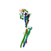

 Sample preparation
 / Beamline: 14.1 / Wavelength: 0.91841 Å
 Processing