 Movie
Movie Controller

[English] 日本語
 Yorodumi
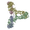
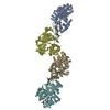
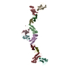
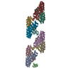
- PDB-1zvo: Semi-extended solution structure of human myeloma immunoglobulin ... -
+ Open data

- Basic information
| Entry | Database: PDB / ID: 1zvo | ||||||
|---|---|---|---|---|---|---|---|
| Title | Semi-extended solution structure of human myeloma immunoglobulin D determined by constrained X-ray scattering | ||||||
 Components Components |
| ||||||
 Keywords Keywords | IMMUNE SYSTEM / IMMUNOGLOBULIN FOLD / ANTIBODY | ||||||
| Function / homology |  Function and homology information Function and homology informationmonocyte activation / positive regulation of interleukin-1 production / CD22 mediated BCR regulation / Fc epsilon receptor (FCERI) signaling / Classical antibody-mediated complement activation / Initial triggering of complement / FCGR activation / Role of LAT2/NTAL/LAB on calcium mobilization / Role of phospholipids in phagocytosis / immunoglobulin complex ...monocyte activation / positive regulation of interleukin-1 production / CD22 mediated BCR regulation / Fc epsilon receptor (FCERI) signaling / Classical antibody-mediated complement activation / Initial triggering of complement / FCGR activation / Role of LAT2/NTAL/LAB on calcium mobilization / Role of phospholipids in phagocytosis / immunoglobulin complex / Scavenging of heme from plasma / antigen binding / FCERI mediated Ca+2 mobilization / FCGR3A-mediated IL10 synthesis / Regulation of Complement cascade / Antigen activates B Cell Receptor (BCR) leading to generation of second messengers / Cell surface interactions at the vascular wall / positive regulation of interleukin-1 beta production / FCGR3A-mediated phagocytosis / FCERI mediated MAPK activation / Regulation of actin dynamics for phagocytic cup formation / Immunoregulatory interactions between a Lymphoid and a non-Lymphoid cell / FCERI mediated NF-kB activation / positive regulation of tumor necrosis factor production / blood microparticle / Potential therapeutics for SARS / adaptive immune response / immune response / : / extracellular region / plasma membrane Similarity search - Function | ||||||
| Biological species |  Homo sapiens (human) Homo sapiens (human) | ||||||
| Method |  SOLUTION SCATTERING / SYNCHROTRON SOLUTION SCATTERING / SYNCHROTRON | ||||||
 Authors Authors | Sun, Z. / Almogren, A. / Furtado, P.B. / Chowdhury, B. / Kerr, M.A. / Perkins, S.J. | ||||||
 Citation Citation | Journal: J.Mol.Biol. / Year: 2005 Title: Semi-extended Solution Structure of Human Myeloma Immunoglobulin D Determined by Constrained X-ray Scattering. Authors: Sun, Z. / Almogren, A. / Furtado, P.B. / Chowdhury, B. / Kerr, M.A. / Perkins, S.J. | ||||||
| History |
|
- Structure visualization
| Structure viewer | Molecule: MolmilJmol/JSmol |
|---|
- Downloads & links
-Download
| PDBx/mmCIF format | 1zvo.cif.gz | 52.9 KB | Display | PDBx/mmCIF format |
|---|---|---|---|---|
| PDB format | pdb1zvo.ent.gz | 30.3 KB | Display | PDB format |
| PDBx/mmJSON format | 1zvo.json.gz | Tree view | PDBx/mmJSON format | |
| Others |  Other downloads Other downloads |
-Validation report
| Arichive directory | https://data.pdbj.org/pub/pdb/validation_reports/zv/1zvoftp://data.pdbj.org/pub/pdb/validation_reports/zv/1zvo | HTTPS FTP |
|---|
-Related structure data
| Related structure data | |
|---|---|
| Similar structure data |


-Links
 PDBj
PDBj


- Assembly
| Deposited unit | 
|
|---|---|
| 1 |
|
-Components
| #1: Antibody | Mass: 22962.377 Da / Num. of mol.: 2 / Source method: isolated from a natural source / Details: Myeloma / Source: (natural) Homo sapiens (human) / References: UniProt: P01700*PLUS#2: Antibody | Mass: 56284.090 Da / Num. of mol.: 2 / Source method: isolated from a natural source / Details: Myeloma / Source: (natural) Homo sapiens (human) / References: UniProt: P0DOX3 |
|---|
-Experimental details
-Experiment
| Experiment | Method: SOLUTION SCATTERING |
|---|
-Data collection
| Diffraction | Mean temperature: 288 K |
|---|---|
| Diffraction source | Source: SYNCHROTRON / Site: ESRF  / Beamline: ID2 / Wavelength: 1 Å / Beamline: ID2 / Wavelength: 1 Å |
| Detector | Detector: CCD / Date: Mar 1, 2002 |
| Radiation | Protocol: SINGLE WAVELENGTH / Monochromatic (M) / Laue (L): M / Scattering type: x-ray |
| Radiation wavelength | Wavelength: 1 Å / Relative weight: 1 |
| Soln scatter | Type: x-ray / Buffer name: 12.5 MM NA PHOSPHATE, 140 MM NACL / Conc. range: 0.30-0.89 / Data analysis software list: SCTPL7, GNOM / Data reduction software list: MULTICCD / Detector type: FRELON CCD CAMERA / Max mean cross sectional radii gyration: 1.24 nm / Max mean cross sectional radii gyration esd: 0.15 nm / Mean guiner radius: 6.94 nm / Mean guiner radius esd: 0.12 nm / Min mean cross sectional radii gyration: 1.93 nm / Min mean cross sectional radii gyration esd: 0.04 nm / Num. of time frames: 1 / Protein length: 1 / Sample pH: 7.2 / Source beamline: ID02 / Source class: Y / Source type: ESRF BEAMLINE ID02 / Temperature: 288 K |
- Processing
| Software | Name: Insight II / Version: II 98.0 / Classification: model building | ||||||||||||
|---|---|---|---|---|---|---|---|---|---|---|---|---|---|
| Refinement step | Cycle: LAST
| ||||||||||||
| Soln scatter model | Method: CONSTRAINED SCATTERING FITTING OF HOMOLOGY MODELS Conformer selection criteria: THE MODELLED SCATTERING CURVES WERE ASSESSED BY CALCULATION OF THE RG AND RXS-1 VALUES IN THE SAME Q RANGES USED IN THE EXPERIMENTAL GUINIER FITS. MODELS WERE THEN ...Conformer selection criteria: THE MODELLED SCATTERING CURVES WERE ASSESSED BY CALCULATION OF THE RG AND RXS-1 VALUES IN THE SAME Q RANGES USED IN THE EXPERIMENTAL GUINIER FITS. MODELS WERE THEN RANKED USING A GOODNESS-OF-FIT R-FACTOR DEFINED BY ANALOGY WITH PROTEIN CRYSTALLOGRAPHY Details: HOMOLOGY MODELS WERE BUILT FOR THE FAB AND FC FRAGMENTS BY TRIAL AND ERROR CONSTRAINED MODELLING. THE POSITIONS OF THE FRAGMENTS WERE DETERMINED BY AN APPROACH THAT COMBINED RANDOMISED HINGE ...Details: HOMOLOGY MODELS WERE BUILT FOR THE FAB AND FC FRAGMENTS BY TRIAL AND ERROR CONSTRAINED MODELLING. THE POSITIONS OF THE FRAGMENTS WERE DETERMINED BY AN APPROACH THAT COMBINED RANDOMISED HINGE PEPTIDE STRUCTURES PRODUCED BY MOLECULAR DYNAMICS SIMULATIONS WITH CURVE-FITTING TO EXPERIMENTAL X-RAY SOLUTION SCATTERING DATA. A SINGLE ARRANGEMENT OF THE FAB AND FC FRAGMENTS IS PRESENTED, WHICH IS REPRESENTATIVE OF A FAMILY OF STRUCTURES THAT FIT THE SCATTERING DATA. MORE DETAILS ON THE MODELLING STRATEGY ARE CONTAINED IN THE PRIMARY REFERENCE. Num. of conformers calculated: 8500 / Num. of conformers submitted: 1 / Representative conformer: 1 / Software author list: ACCELRYS Software list: INSIGHT II, HOMOLOGY, DISCOVERY, BIOPOLYMER, DELPHI, O, SCTPL7, GNOM |
