 Movie
Movie Controller


+ Open data

- Basic information
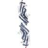
| Entry | Database: PDB / ID: 4aqt | |||||||||
|---|---|---|---|---|---|---|---|---|---|---|
| Title | Laminin gamma1 LN-LE1-2 structure | |||||||||
 Components Components | LAMININ SUBUNIT GAMMA-1 | |||||||||
 Keywords Keywords | CELL ADHESION | |||||||||
| Function / homology |  Function and homology information Function and homology informationlaminin-111 trimer / laminin-511 trimer / tissue morphogenesis / hair follicle cell proliferation / regulation of basement membrane organization / hemidesmosome assembly / glycosphingolipid binding / positive regulation of integrin-mediated signaling pathway / tissue development / hair cell differentiation ...laminin-111 trimer / laminin-511 trimer / tissue morphogenesis / hair follicle cell proliferation / regulation of basement membrane organization / hemidesmosome assembly / glycosphingolipid binding / positive regulation of integrin-mediated signaling pathway / tissue development / hair cell differentiation / protein complex involved in cell-matrix adhesion / positive regulation of skeletal muscle acetylcholine-gated channel clustering / hair follicle morphogenesis / positive regulation of muscle cell differentiation / extracellular matrix structural constituent / basement membrane / extracellular matrix disassembly / synaptic cleft / substrate adhesion-dependent cell spreading / positive regulation of cell adhesion / axon guidance / animal organ morphogenesis / neuromuscular junction / neuron projection development / cell migration / chromatin organization / extracellular matrix / protein-containing complex assembly / gene expression / cell adhesion / receptor ligand activity / extracellular region Similarity search - Function | |||||||||
| Biological species |  | |||||||||
| Method |  X-RAY DIFFRACTION / SYNCHROTRON / MOLECULAR REPLACEMENT / Resolution: 3.2 Å X-RAY DIFFRACTION / SYNCHROTRON / MOLECULAR REPLACEMENT / Resolution: 3.2 Å | |||||||||
 Authors Authors | Carafoli, F. / Hussain, S. / Hohenester, E. | |||||||||
 Citation Citation | Journal: Plos One / Year: 2012 Title: Crystal Structures of the Network-Forming Short-Arm Tips of the Laminin Beta1 and Gamma1 Chains. Authors: Carafoli, F. / Hussain, S. / Hohenester, E. | |||||||||
| History |
|
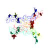
- Structure visualization
| Structure viewer | Molecule: MolmilJmol/JSmol |
|---|
- Downloads & links
-Download
| PDBx/mmCIF format | 4aqt.cif.gz | 85.8 KB | Display | PDBx/mmCIF format |
|---|---|---|---|---|
| PDB format | pdb4aqt.ent.gz | 62.7 KB | Display | PDB format |
| PDBx/mmJSON format | 4aqt.json.gz | Tree view | PDBx/mmJSON format | |
| Others |  Other downloads Other downloads |
-Validation report
| Arichive directory | https://data.pdbj.org/pub/pdb/validation_reports/aq/4aqtftp://data.pdbj.org/pub/pdb/validation_reports/aq/4aqt | HTTPS FTP |
|---|
-Related structure data
| Related structure data | 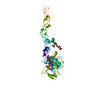 4aqsC  2y38S C: citing same article ( S: Starting model for refinement |
|---|---|
| Similar structure data |


-Links
 PDBj
PDBj

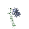
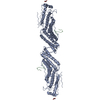
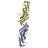
- Assembly
| Deposited unit | 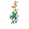
| ||||||||
|---|---|---|---|---|---|---|---|---|---|
| 1 | 
| ||||||||
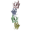
| Unit cell |
|
-Components
| #1: Protein | Mass: 41976.402 Da / Num. of mol.: 1 / Fragment: LN-LE1-2 FRAGMENT, RESIDUES 33-395 Source method: isolated from a genetically manipulated source Source: (gene. exp.)  HOMO SAPIENS (human) / References: UniProt: P02468 HOMO SAPIENS (human) / References: UniProt: P02468 |
|---|---|
| #2: Polysaccharide | beta-D-mannopyranose-(1-4)-2-acetamido-2-deoxy-beta-D-glucopyranose-(1-4)-2-acetamido-2-deoxy-beta- ...beta-D-mannopyranose-(1-4)-2-acetamido-2-deoxy-beta-D-glucopyranose-(1-4)-2-acetamido-2-deoxy-beta-D-glucopyranose Source method: isolated from a genetically manipulated source |
| #3: Chemical | ChemComp-CA /   Mass: 40.078 Da / Num. of mol.: 1 / Source method: obtained synthetically / Formula: Ca Mass: 40.078 Da / Num. of mol.: 1 / Source method: obtained synthetically / Formula: Ca |
| #4: Sugar | ChemComp-NAG /   Type: D-saccharide, beta linking / Mass: 221.208 Da / Num. of mol.: 1 Type: D-saccharide, beta linking / Mass: 221.208 Da / Num. of mol.: 1Source method: isolated from a genetically manipulated source Formula: C8H15NO6 |
| Has protein modification | Y |
-Experimental details
-Experiment
| Experiment | Method: X-RAY DIFFRACTION / Number of used crystals: 1 |
|---|
- Sample preparation
| Crystal | Density Matthews: 4.43 Å3/Da / Density % sol: 72 % Description: THE DIFFRACTION LIMIT OF THE CRYSTAL WAS ANISOTROPIC AND LIMITED TO 3.8 A ALONG THE C AXIS. |
|---|---|
| Crystal grow | pH: 6.5 / Details: pH 6.5 |
-Data collection
| Diffraction | Mean temperature: 100 K |
|---|---|
| Diffraction source | Source: SYNCHROTRON / Site: Diamond  / Beamline: I04 / Wavelength: 0.9763 / Beamline: I04 / Wavelength: 0.9763 |
| Detector | Type: ADSC CCD / Detector: CCD / Date: May 13, 2010 |
| Radiation | Protocol: SINGLE WAVELENGTH / Monochromatic (M) / Laue (L): M / Scattering type: x-ray |
| Radiation wavelength | Wavelength: 0.9763 Å / Relative weight: 1 |
| Reflection | Resolution: 3.17→50 Å / Num. obs: 12556 / % possible obs: 99.4 % / Observed criterion σ(I): 0 / Redundancy: 11 % / Biso Wilson estimate: 83.4 Å2 / Rmerge(I) obs: 0.1 / Net I/σ(I): 18.6 |
| Reflection shell | Resolution: 3.17→3.25 Å / Redundancy: 11.2 % / Rmerge(I) obs: 0.67 / Mean I/σ(I) obs: 4.5 / % possible all: 100 |
- Processing
| Software |
| ||||||||||||||||||||||||||||||||||||||||||||||||||||||||||||||||||||||||||||||||
|---|---|---|---|---|---|---|---|---|---|---|---|---|---|---|---|---|---|---|---|---|---|---|---|---|---|---|---|---|---|---|---|---|---|---|---|---|---|---|---|---|---|---|---|---|---|---|---|---|---|---|---|---|---|---|---|---|---|---|---|---|---|---|---|---|---|---|---|---|---|---|---|---|---|---|---|---|---|---|---|---|---|
| Refinement | Method to determine structure: MOLECULAR REPLACEMENT Starting model: PDB ENTRY 2Y38 Resolution: 3.2→50 Å / Data cutoff high absF: 10000 / Data cutoff low absF: 0 / Isotropic thermal model: RESTRAINED INDIVIDUAL / Cross valid method: THROUGHOUT / σ(F): 0 Details: BECAUSE OF THE ANISOTROPIC DIFFRACTION LIMIT, THE DATA WERE TRUNCATED AT 3.8 A ALONG THE C-AXIS AND AT 3.2 A ALONG THE A- AND B-AXES USING THE DIFFRACTION ANISOTROPY SERVER
| ||||||||||||||||||||||||||||||||||||||||||||||||||||||||||||||||||||||||||||||||
| Solvent computation | Solvent model: FLAT / Bsol: 18.1433 Å2 / ksol: 0.28 e/Å3 | ||||||||||||||||||||||||||||||||||||||||||||||||||||||||||||||||||||||||||||||||
| Displacement parameters | Biso mean: 63.6 Å2
| ||||||||||||||||||||||||||||||||||||||||||||||||||||||||||||||||||||||||||||||||
| Refinement step | Cycle: LAST / Resolution: 3.2→50 Å
| ||||||||||||||||||||||||||||||||||||||||||||||||||||||||||||||||||||||||||||||||
| Refine LS restraints |
| ||||||||||||||||||||||||||||||||||||||||||||||||||||||||||||||||||||||||||||||||
| Xplor file |
|
