 Movie
Movie Controller

[English] 日本語
 Yorodumi
- PDB-1klo: CRYSTAL STRUCTURE OF THREE CONSECUTIVE LAMININ-TYPE EPIDERMAL GRO... -
+ Open data

- Basic information
| Entry | Database: PDB / ID: 1klo | ||||||
|---|---|---|---|---|---|---|---|
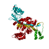
| Title | CRYSTAL STRUCTURE OF THREE CONSECUTIVE LAMININ-TYPE EPIDERMAL GROWTH FACTOR-LIKE (LE) MODULES OF LAMININ GAMMA1 CHAIN HARBORING THE NIDOGEN BINDING SITE | ||||||
 Components Components | LAMININ | ||||||
 Keywords Keywords | GLYCOPROTEIN | ||||||
| Function / homology |  Function and homology information Function and homology informationlaminin-111 trimer / laminin-511 trimer / tissue morphogenesis / hair follicle cell proliferation / regulation of basement membrane organization / hemidesmosome assembly / glycosphingolipid binding / positive regulation of integrin-mediated signaling pathway / tissue development / hair cell differentiation ...laminin-111 trimer / laminin-511 trimer / tissue morphogenesis / hair follicle cell proliferation / regulation of basement membrane organization / hemidesmosome assembly / glycosphingolipid binding / positive regulation of integrin-mediated signaling pathway / tissue development / hair cell differentiation / protein complex involved in cell-matrix adhesion / positive regulation of skeletal muscle acetylcholine-gated channel clustering / hair follicle morphogenesis / positive regulation of muscle cell differentiation / extracellular matrix structural constituent / basement membrane / extracellular matrix disassembly / synaptic cleft / substrate adhesion-dependent cell spreading / positive regulation of cell adhesion / axon guidance / animal organ morphogenesis / neuromuscular junction / neuron projection development / cell migration / chromatin organization / extracellular matrix / protein-containing complex assembly / gene expression / cell adhesion / receptor ligand activity / extracellular region Similarity search - Function | ||||||
| Biological species |  | ||||||
| Method |  X-RAY DIFFRACTION / Resolution: 2.1 Å X-RAY DIFFRACTION / Resolution: 2.1 Å | ||||||
 Authors Authors | Stetefeld, J. / Mayer, U. / Timpl, R. / Huber, R. | ||||||
 Citation Citation | Journal: J.Mol.Biol. / Year: 1996 Title: Crystal structure of three consecutive laminin-type epidermal growth factor-like (LE) modules of laminin gamma1 chain harboring the nidogen binding site. Authors: Stetefeld, J. / Mayer, U. / Timpl, R. / Huber, R. #1: Journal: Embo J. / Year: 1994Title: Two Non-Contiguous Regions Contribute to Nidogen Binding to a Single Egf-Like Motif of the Laminin Gamma 1 Chain Authors: Poschl, E. / Fox, J.W. / Block, D. / Mayer, U. / Timpl, R. #2: Journal: Embo J. / Year: 1993Title: A Single Egf-Like Motif of Laminin is Responsible for High Affinity Nidogen Binding Authors: Mayer, U. / Nischt, R. / Poschl, E. / Mann, K. / Fukuda, K. / Gerl, M. / Yamada, Y. / Timpl, R. | ||||||
| History |
|
- Structure visualization
| Structure viewer | Molecule: MolmilJmol/JSmol |
|---|
- Downloads & links
-Download
| PDBx/mmCIF format | 1klo.cif.gz | 54.4 KB | Display | PDBx/mmCIF format |
|---|---|---|---|---|
| PDB format | pdb1klo.ent.gz | 39.4 KB | Display | PDB format |
| PDBx/mmJSON format | 1klo.json.gz | Tree view | PDBx/mmJSON format | |
| Others |  Other downloads Other downloads |
-Validation report
| Arichive directory | https://data.pdbj.org/pub/pdb/validation_reports/kl/1kloftp://data.pdbj.org/pub/pdb/validation_reports/kl/1klo | HTTPS FTP |
|---|
-Related structure data
| Similar structure data |
|---|


-Links
 PDBj
PDBj

- Assembly
| Deposited unit | 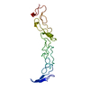
| ||||||||
|---|---|---|---|---|---|---|---|---|---|
| 1 | 
| ||||||||
| Unit cell |
|
-Components
| #1: Protein | Mass: 17141.363 Da / Num. of mol.: 1 Fragment: GAMMA-1 CHAIN, THREE CONSECUTIVE LAMININ-TYPE EPIDERMAL GROWTH FACTOR-LIKE (LE) MODULES Source method: isolated from a natural source / Source: (natural) |
|---|---|
| #2: Water | ChemComp-HOH /  Mass: 18.015 Da / Num. of mol.: 119 / Source method: isolated from a natural source / Formula: H2O Mass: 18.015 Da / Num. of mol.: 119 / Source method: isolated from a natural source / Formula: H2O |
| Has protein modification | Y |
-Experimental details
-Experiment
| Experiment | Method: X-RAY DIFFRACTION |
|---|
- Sample preparation
| Crystal | Density Matthews: 4.33 Å3/Da / Density % sol: 71.59 % | ||||||||||||||||||||||||||||||||||||||||||||||||||||||||
|---|---|---|---|---|---|---|---|---|---|---|---|---|---|---|---|---|---|---|---|---|---|---|---|---|---|---|---|---|---|---|---|---|---|---|---|---|---|---|---|---|---|---|---|---|---|---|---|---|---|---|---|---|---|---|---|---|---|
| Crystal | *PLUS Density % sol: 4.1 % | ||||||||||||||||||||||||||||||||||||||||||||||||||||||||
| Crystal grow | *PLUS Temperature: 22 ℃ / pH: 7 / Method: vapor diffusion, hanging drop | ||||||||||||||||||||||||||||||||||||||||||||||||||||||||
| Components of the solutions | *PLUS
|
-Data collection
| Radiation | Scattering type: x-ray |
|---|---|
| Radiation wavelength | Relative weight: 1 |
| Reflection | *PLUS Highest resolution: 2.1 Å / Num. obs: 17012 / % possible obs: 91.4 % / Num. measured all: 29004 / Rmerge(I) obs: 0.07 |
- Processing
| Software |
| ||||||||||||||||||||||||||||||||||||||||||||||||||||||||||||
|---|---|---|---|---|---|---|---|---|---|---|---|---|---|---|---|---|---|---|---|---|---|---|---|---|---|---|---|---|---|---|---|---|---|---|---|---|---|---|---|---|---|---|---|---|---|---|---|---|---|---|---|---|---|---|---|---|---|---|---|---|---|
| Refinement | Resolution: 2.1→6 Å / σ(F): 0 /
| ||||||||||||||||||||||||||||||||||||||||||||||||||||||||||||
| Refinement step | Cycle: LAST / Resolution: 2.1→6 Å
| ||||||||||||||||||||||||||||||||||||||||||||||||||||||||||||
| Refine LS restraints |
| ||||||||||||||||||||||||||||||||||||||||||||||||||||||||||||
| Refinement | *PLUS | ||||||||||||||||||||||||||||||||||||||||||||||||||||||||||||
| Solvent computation | *PLUS | ||||||||||||||||||||||||||||||||||||||||||||||||||||||||||||
| Displacement parameters | *PLUS | ||||||||||||||||||||||||||||||||||||||||||||||||||||||||||||
| Refine LS restraints | *PLUS
|
