 Movie
Movie Controller


+ Open data

- Basic information
| Entry | Database: PDB / ID: 1qb3 | ||||||
|---|---|---|---|---|---|---|---|
| Title | CRYSTAL STRUCTURE OF THE CELL CYCLE REGULATORY PROTEIN CKS1 | ||||||
 Components Components | CYCLIN-DEPENDENT KINASES REGULATORY SUBUNIT | ||||||
 Keywords Keywords | CELL CYCLE / CELL CYCLE MUTAGENESIS DOMAIN SWAPPING / CYCLIN-DEPENDENT KINASE | ||||||
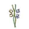
| Function / homology |  Function and homology information Function and homology informationregulation of cell cycle G1/S phase transition / cyclin-dependent protein serine/threonine kinase activator activity / SCF ubiquitin ligase complex / protein kinase activator activity / cyclin-dependent protein kinase holoenzyme complex / regulation of mitotic cell cycle / ubiquitin binding / histone binding / cell division / regulation of transcription by RNA polymerase II ...regulation of cell cycle G1/S phase transition / cyclin-dependent protein serine/threonine kinase activator activity / SCF ubiquitin ligase complex / protein kinase activator activity / cyclin-dependent protein kinase holoenzyme complex / regulation of mitotic cell cycle / ubiquitin binding / histone binding / cell division / regulation of transcription by RNA polymerase II / protein kinase binding / positive regulation of transcription by RNA polymerase II / nucleus / cytoplasm Similarity search - Function | ||||||
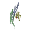
| Biological species |  | ||||||
| Method |  X-RAY DIFFRACTION / SYNCHROTRON / Resolution: 3 Å X-RAY DIFFRACTION / SYNCHROTRON / Resolution: 3 Å | ||||||
 Authors Authors | Bourne, Y. / Watson, M.H. / Arvai, A.S. / Bernstein, S.L. / Reed, S.I. / Tainer, J.A. | ||||||
 Citation Citation | Journal: Structure Fold.Des. / Year: 2000 Title: Crystal structure and mutational analysis of the Saccharomyces cerevisiae cell cycle regulatory protein Cks1: implications for domain swapping, anion binding and protein interactions. Authors: Bourne, Y. / Watson, M.H. / Arvai, A.S. / Bernstein, S.L. / Reed, S.I. / Tainer, J.A. | ||||||
| History |
|
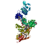
- Structure visualization
| Structure viewer | Molecule: MolmilJmol/JSmol |
|---|
- Downloads & links
-Download
| PDBx/mmCIF format | 1qb3.cif.gz | 83.3 KB | Display | PDBx/mmCIF format |
|---|---|---|---|---|
| PDB format | pdb1qb3.ent.gz | 63.4 KB | Display | PDB format |
| PDBx/mmJSON format | 1qb3.json.gz | Tree view | PDBx/mmJSON format | |
| Others |  Other downloads Other downloads |
-Validation report
| Arichive directory | https://data.pdbj.org/pub/pdb/validation_reports/qb/1qb3ftp://data.pdbj.org/pub/pdb/validation_reports/qb/1qb3 | HTTPS FTP |
|---|
-Related structure data
| Similar structure data |
|---|


-Links
 PDBj
PDBj
- Assembly
| Deposited unit | 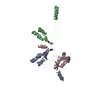
| ||||||||
|---|---|---|---|---|---|---|---|---|---|
| 1 | 
| ||||||||
| 2 | 
| ||||||||
| 3 | 
| ||||||||
| 4 | 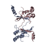
| ||||||||
| 5 | 
| ||||||||
| Unit cell |
|
-Components
| #1: Protein | Mass: 17816.715 Da / Num. of mol.: 3 Source method: isolated from a genetically manipulated source Source: (gene. exp.) Plasmid: PBR322 / Production host:  #2: Water | ChemComp-HOH / |  Mass: 18.015 Da / Num. of mol.: 24 / Source method: isolated from a natural source / Formula: H2O Mass: 18.015 Da / Num. of mol.: 24 / Source method: isolated from a natural source / Formula: H2OHas protein modification | Y | |
|---|
-Experimental details
-Experiment
| Experiment | Method: X-RAY DIFFRACTION / Number of used crystals: 1 |
|---|
- Sample preparation
| Crystal | Density Matthews: 2.51 Å3/Da / Density % sol: 50.93 % | ||||||||||||||||||||||||||||||
|---|---|---|---|---|---|---|---|---|---|---|---|---|---|---|---|---|---|---|---|---|---|---|---|---|---|---|---|---|---|---|---|
| Crystal grow | Temperature: 293 K / Method: vapor diffusion, sitting drop / pH: 7 Details: 20% PEG 4K, 5% NASCN AND 0.1 M IMIDAZOLE/MALATE, pH 7.0, VAPOR DIFFUSION, SITTING DROP, temperature 293K | ||||||||||||||||||||||||||||||
| Crystal | *PLUS Density % sol: 51 % | ||||||||||||||||||||||||||||||
| Crystal grow | *PLUS Temperature: 20 ℃ / Method: vapor diffusion | ||||||||||||||||||||||||||||||
| Components of the solutions | *PLUS
|
-Data collection
| Diffraction | Mean temperature: 100 K |
|---|---|
| Diffraction source | Source: SYNCHROTRON / Site: SSRL  / Beamline: BL7-1 / Wavelength: 1.08 / Beamline: BL7-1 / Wavelength: 1.08 |
| Detector | Type: MARRESEARCH / Detector: IMAGE PLATE / Date: Oct 20, 1995 |
| Radiation | Protocol: SINGLE WAVELENGTH / Monochromatic (M) / Laue (L): M / Scattering type: x-ray |
| Radiation wavelength | Wavelength: 1.08 Å / Relative weight: 1 |
| Reflection | Resolution: 3→12 Å / Num. all: 10366 / Num. obs: 10218 / % possible obs: 91 % / Observed criterion σ(F): 0 / Redundancy: 12.6 % / Biso Wilson estimate: 57 Å2 / Rmerge(I) obs: 0.094 / Net I/σ(I): 7.4 |
| Reflection shell | Resolution: 3→3.1 Å / Redundancy: 11.7 % / Rmerge(I) obs: 0.405 / % possible all: 93.7 |
| Reflection | *PLUS Num. obs: 10366 / Num. measured all: 243609 |
- Processing
| Software |
| ||||||||||||||||||||||||||||||||||||||||||||||||||||||||||||
|---|---|---|---|---|---|---|---|---|---|---|---|---|---|---|---|---|---|---|---|---|---|---|---|---|---|---|---|---|---|---|---|---|---|---|---|---|---|---|---|---|---|---|---|---|---|---|---|---|---|---|---|---|---|---|---|---|---|---|---|---|---|
| Refinement | Resolution: 3→12 Å / σ(F): 0 / Stereochemistry target values: ENGH & HUBER
| ||||||||||||||||||||||||||||||||||||||||||||||||||||||||||||
| Refinement step | Cycle: LAST / Resolution: 3→12 Å
| ||||||||||||||||||||||||||||||||||||||||||||||||||||||||||||
| Refine LS restraints |
| ||||||||||||||||||||||||||||||||||||||||||||||||||||||||||||
| Software | *PLUS Name: CNS / Classification: refinement | ||||||||||||||||||||||||||||||||||||||||||||||||||||||||||||
| Refinement | *PLUS Highest resolution: 3 Å / Lowest resolution: 12 Å / σ(F): 0 / % reflection Rfree: 5 % / Rfactor obs: 0.216 / Num. reflection obs: 10366 | ||||||||||||||||||||||||||||||||||||||||||||||||||||||||||||
| Solvent computation | *PLUS | ||||||||||||||||||||||||||||||||||||||||||||||||||||||||||||
| Displacement parameters | *PLUS | ||||||||||||||||||||||||||||||||||||||||||||||||||||||||||||
| Refine LS restraints | *PLUS
|
