 Movie
Movie Controller

[English] 日本語
 Yorodumi
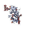
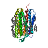
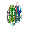
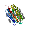
- PDB-4dvn: Crystal structure of the glycoprotein Erns from the pestivirus BV... -
+ Open data

- Basic information
| Entry | Database: PDB / ID: 4dvn | |||||||||
|---|---|---|---|---|---|---|---|---|---|---|
| Title | Crystal structure of the glycoprotein Erns from the pestivirus BVDV-1 in complex with 2'-UMP | |||||||||
 Components Components | E(rns) glycoprotein | |||||||||
 Keywords Keywords | VIRAL PROTEIN / virus glycoprotein / T2 Ribonuclease | |||||||||
| Function / homology |  Function and homology information Function and homology informationpestivirus NS3 polyprotein peptidase / ribonuclease T2 / serine-type exopeptidase activity / ribonuclease T2 activity / nucleoside-triphosphate phosphatase / channel activity / monoatomic ion transmembrane transport / clathrin-dependent endocytosis of virus by host cell / symbiont-mediated suppression of host cytoplasmic pattern recognition receptor signaling pathway via inhibition of IRF3 activity / Hydrolases; Acting on peptide bonds (peptidases); Cysteine endopeptidases ...pestivirus NS3 polyprotein peptidase / ribonuclease T2 / serine-type exopeptidase activity / ribonuclease T2 activity / nucleoside-triphosphate phosphatase / channel activity / monoatomic ion transmembrane transport / clathrin-dependent endocytosis of virus by host cell / symbiont-mediated suppression of host cytoplasmic pattern recognition receptor signaling pathway via inhibition of IRF3 activity / Hydrolases; Acting on peptide bonds (peptidases); Cysteine endopeptidases / RNA helicase activity / viral protein processing / host cell endoplasmic reticulum membrane / RNA helicase / serine-type endopeptidase activity / symbiont-mediated activation of host autophagy / RNA-directed RNA polymerase / cysteine-type endopeptidase activity / viral RNA genome replication / RNA-directed RNA polymerase activity / fusion of virus membrane with host endosome membrane / virion attachment to host cell / GTP binding / virion membrane / ATP hydrolysis activity / proteolysis / RNA binding / ATP binding Similarity search - Function | |||||||||
| Biological species |  Bovine viral diarrhea virus Bovine viral diarrhea virus | |||||||||
| Method |  X-RAY DIFFRACTION / SYNCHROTRON / MOLECULAR REPLACEMENT / molecular replacement / Resolution: 2.38 Å X-RAY DIFFRACTION / SYNCHROTRON / MOLECULAR REPLACEMENT / molecular replacement / Resolution: 2.38 Å | |||||||||
 Authors Authors | Krey, T. / Bontems, F. / Vonrhein, C. / Vaney, M.-C. / Bricogne, G. / Ruemenapf, T. / Rey, F.A. | |||||||||
 Citation Citation | Journal: Structure / Year: 2012 Title: Crystal Structure of the Pestivirus Envelope Glycoprotein E(rns) and Mechanistic Analysis of Its Ribonuclease Activity. Authors: Krey, T. / Bontems, F. / Vonrhein, C. / Vaney, M.C. / Bricogne, G. / Rumenapf, T. / Rey, F.A. | |||||||||
| History |
|
- Structure visualization
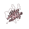
| Structure viewer | Molecule: MolmilJmol/JSmol |
|---|
- Downloads & links
-Download
| PDBx/mmCIF format | 4dvn.cif.gz | 92.3 KB | Display | PDBx/mmCIF format |
|---|---|---|---|---|
| PDB format | pdb4dvn.ent.gz | 70.7 KB | Display | PDB format |
| PDBx/mmJSON format | 4dvn.json.gz | Tree view | PDBx/mmJSON format | |
| Others |  Other downloads Other downloads |
-Validation report
| Arichive directory | https://data.pdbj.org/pub/pdb/validation_reports/dv/4dvnftp://data.pdbj.org/pub/pdb/validation_reports/dv/4dvn | HTTPS FTP |
|---|
-Related structure data
| Related structure data |  4dvkSC  4dvlC  4dw3C  4dw4C  4dw5C  4dw7C  4dwaC  4dwcC S: Starting model for refinement C: citing same article ( |
|---|---|
| Similar structure data |


-Links
 PDBj
PDBj
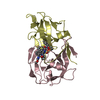
- Assembly
| Deposited unit | 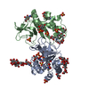
| ||||||||
|---|---|---|---|---|---|---|---|---|---|
| 1 | 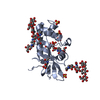
| ||||||||
| 2 | 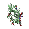
| ||||||||
| Unit cell |
| ||||||||
| Components on special symmetry positions |
|
-Components
-Protein , 1 types, 2 molecules AB
| #1: Protein | Mass: 19005.492 Da / Num. of mol.: 2 / Fragment: N-terminal fragment Source method: isolated from a genetically manipulated source Source: (gene. exp.) Bovine viral diarrhea virus / Strain: CP7 / Gene: Erns / Plasmid: pMT-BIP-based / Production host:  |
|---|
-Sugars , 5 types, 9 molecules 
| #2: Polysaccharide | alpha-D-mannopyranose-(1-3)-[beta-D-mannopyranose-(1-6)]beta-D-mannopyranose-(1-4)-2-acetamido-2- ...alpha-D-mannopyranose-(1-3)-[beta-D-mannopyranose-(1-6)]beta-D-mannopyranose-(1-4)-2-acetamido-2-deoxy-beta-D-glucopyranose-(1-4)-2-acetamido-2-deoxy-beta-D-glucopyranose Source method: isolated from a genetically manipulated source |
|---|---|
| #3: Polysaccharide | beta-D-mannopyranose-(1-3)-[alpha-D-mannopyranose-(1-6)]beta-D-mannopyranose-(1-4)-2-acetamido-2- ...beta-D-mannopyranose-(1-3)-[alpha-D-mannopyranose-(1-6)]beta-D-mannopyranose-(1-4)-2-acetamido-2-deoxy-beta-D-glucopyranose-(1-4)-2-acetamido-2-deoxy-beta-D-glucopyranose Source method: isolated from a genetically manipulated source |
| #4: Polysaccharide | alpha-D-mannopyranose-(1-3)-[alpha-D-mannopyranose-(1-6)]beta-D-mannopyranose-(1-4)-2-acetamido-2- ...alpha-D-mannopyranose-(1-3)-[alpha-D-mannopyranose-(1-6)]beta-D-mannopyranose-(1-4)-2-acetamido-2-deoxy-beta-D-glucopyranose-(1-4)-2-acetamido-2-deoxy-beta-D-glucopyranose Source method: isolated from a genetically manipulated source |
| #5: Polysaccharide | 2-acetamido-2-deoxy-beta-D-glucopyranose-(1-4)-2-acetamido-2-deoxy-beta-D-glucopyranose Source method: isolated from a genetically manipulated source |
| #6: Sugar | ChemComp-NAG /  Type: D-saccharide, beta linking / Mass: 221.208 Da / Num. of mol.: 5 Type: D-saccharide, beta linking / Mass: 221.208 Da / Num. of mol.: 5Source method: isolated from a genetically manipulated source Formula: C8H15NO6 |
-Non-polymers , 3 types, 171 molecules 

| #7: Chemical | ChemComp-SO4 /  Mass: 96.063 Da / Num. of mol.: 7 / Source method: obtained synthetically / Formula: SO4 Mass: 96.063 Da / Num. of mol.: 7 / Source method: obtained synthetically / Formula: SO4#8: Chemical |  Type: RNA linking / Mass: 324.181 Da / Num. of mol.: 3 / Source method: obtained synthetically / Formula: C9H13N2O9P Type: RNA linking / Mass: 324.181 Da / Num. of mol.: 3 / Source method: obtained synthetically / Formula: C9H13N2O9P#9: Water | ChemComp-HOH / | Mass: 18.015 Da / Num. of mol.: 161 / Source method: isolated from a natural source / Formula: H2O |
|---|
-Details
| Has protein modification | Y |
|---|
-Experimental details
-Experiment
| Experiment | Method: X-RAY DIFFRACTION / Number of used crystals: 1 |
|---|
- Sample preparation
| Crystal | Density Matthews: 3.17 Å3/Da / Density % sol: 61.17 % |
|---|---|
| Crystal grow | Temperature: 298 K / Method: vapor diffusion / pH: 4.6 Details: 33% PEG2000 MME, 100mM Na-Acetate, 140mM (NH4)2SO4, 50mM KH2PO4, pH 4.6, vapor diffusion, temperature 298K |
-Data collection
| Diffraction | Mean temperature: 110 K |
|---|---|
| Diffraction source | Source: SYNCHROTRON / Site: ESRF  / Beamline: ID14-1 / Wavelength: 0.9334 Å / Beamline: ID14-1 / Wavelength: 0.9334 Å |
| Detector | Type: ADSC QUANTUM 210 / Detector: CCD / Date: Sep 1, 2008 |
| Radiation | Monochromator: Diamond (001) / Protocol: SINGLE WAVELENGTH / Monochromatic (M) / Laue (L): M / Scattering type: x-ray |
| Radiation wavelength | Wavelength: 0.9334 Å / Relative weight: 1 |
| Reflection | Resolution: 2.38→47.41 Å / Num. all: 19827 / Num. obs: 17289 / % possible obs: 87.2 % / Observed criterion σ(F): 0 / Observed criterion σ(I): -3 / Biso Wilson estimate: 37.69 Å2 |
| Reflection shell | Resolution: 2.38→2.51 Å / % possible all: 64.9 |
-Phasing
| Phasing | Method: molecular replacement |
|---|
- Processing
| Software |
| ||||||||||||||||||||||||||||||||||||||||||||||||||||||||||||||||||||||||||||||||||||||||||||||||||||||||||||||||||
|---|---|---|---|---|---|---|---|---|---|---|---|---|---|---|---|---|---|---|---|---|---|---|---|---|---|---|---|---|---|---|---|---|---|---|---|---|---|---|---|---|---|---|---|---|---|---|---|---|---|---|---|---|---|---|---|---|---|---|---|---|---|---|---|---|---|---|---|---|---|---|---|---|---|---|---|---|---|---|---|---|---|---|---|---|---|---|---|---|---|---|---|---|---|---|---|---|---|---|---|---|---|---|---|---|---|---|---|---|---|---|---|---|---|---|---|
| Refinement | Method to determine structure: MOLECULAR REPLACEMENT Starting model: PDB entry 4DVK Resolution: 2.38→22.85 Å / Cor.coef. Fo:Fc: 0.9044 / Cor.coef. Fo:Fc free: 0.8884 / Occupancy max: 1 / Occupancy min: 0.5 / Cross valid method: THROUGHOUT / σ(F): 0 / Stereochemistry target values: Engh & Huber
| ||||||||||||||||||||||||||||||||||||||||||||||||||||||||||||||||||||||||||||||||||||||||||||||||||||||||||||||||||
| Displacement parameters | Biso mean: 33.59 Å2
| ||||||||||||||||||||||||||||||||||||||||||||||||||||||||||||||||||||||||||||||||||||||||||||||||||||||||||||||||||
| Refine analyze | Luzzati coordinate error obs: 0.289 Å | ||||||||||||||||||||||||||||||||||||||||||||||||||||||||||||||||||||||||||||||||||||||||||||||||||||||||||||||||||
| Refinement step | Cycle: LAST / Resolution: 2.38→22.85 Å
| ||||||||||||||||||||||||||||||||||||||||||||||||||||||||||||||||||||||||||||||||||||||||||||||||||||||||||||||||||
| Refine LS restraints |
| ||||||||||||||||||||||||||||||||||||||||||||||||||||||||||||||||||||||||||||||||||||||||||||||||||||||||||||||||||
| LS refinement shell | Resolution: 2.38→2.52 Å / Total num. of bins used: 9
|
