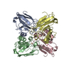
 Movie
Movie Controller


+ Open data

- Basic information
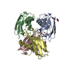
| Entry | Database: PDB / ID: 1vlx | ||||||
|---|---|---|---|---|---|---|---|
| Title | STRUCTURE OF ELECTRON TRANSFER (COBALT-PROTEIN) | ||||||
 Components Components | AZURIN | ||||||
 Keywords Keywords | ELECTRON TRANSPORT / COPPER / PERIPLASMIC | ||||||
| Function / homology |  Function and homology information Function and homology informationtransition metal ion binding / electron transfer activity / periplasmic space / copper ion binding / zinc ion binding / identical protein binding / plasma membrane Similarity search - Function | ||||||
| Biological species |   Pseudomonas aeruginosa (bacteria) Pseudomonas aeruginosa (bacteria) | ||||||
| Method |  X-RAY DIFFRACTION / ISOMORPHOUS REPLACEMENT / Resolution: 1.9 Å X-RAY DIFFRACTION / ISOMORPHOUS REPLACEMENT / Resolution: 1.9 Å | ||||||
 Authors Authors | Bonander, N. / Vanngard, T. / Tsai, L.-C. / Langer, V. / Nar, H. / Sjolin, L. | ||||||
 Citation Citation | Journal: Proteins / Year: 1997 Title: The metal site of Pseudomonas aeruginosa azurin, revealed by a crystal structure determination of the Co(II) derivative and Co-EPR spectroscopy. Authors: Bonander, N. / Vanngard, T. / Tsai, L.C. / Langer, V. / Nar, H. / Sjolin, L. #1: Journal: To be PublishedTitle: X-Ray Structure Determination and Characterization of the Pseudomonas Aeruginosa Azurin Mutant met121Glu Authors: Karlsson, B.G. / Tsai, L.-C. / Nar, H. / Sanders-Loehr, J. / Bonander, N. / Langer, V. / Sjoelin, L. | ||||||
| History |
|
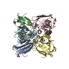
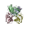
- Structure visualization
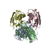
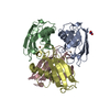
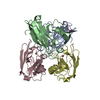
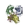
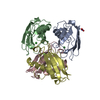
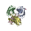
| Structure viewer | Molecule: MolmilJmol/JSmol |
|---|
- Downloads & links
-Download
| PDBx/mmCIF format | 1vlx.cif.gz | 114.8 KB | Display | PDBx/mmCIF format |
|---|---|---|---|---|
| PDB format | pdb1vlx.ent.gz | 88.9 KB | Display | PDB format |
| PDBx/mmJSON format | 1vlx.json.gz | Tree view | PDBx/mmJSON format | |
| Others |  Other downloads Other downloads |
-Validation report
| Arichive directory | https://data.pdbj.org/pub/pdb/validation_reports/vl/1vlxftp://data.pdbj.org/pub/pdb/validation_reports/vl/1vlx | HTTPS FTP |
|---|
-Related structure data
| Similar structure data |
|---|


-Links
 PDBj
PDBj
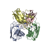
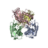
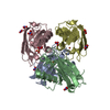
- Assembly
| Deposited unit | 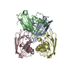
| ||||||||
|---|---|---|---|---|---|---|---|---|---|
| 1 |
| ||||||||
| Unit cell |
|
-Components
| #1: Protein | Mass: 13961.799 Da / Num. of mol.: 4 / Source method: isolated from a natural source / Details: COBALT FORM / Source: (natural) Pseudomonas aeruginosa (bacteria) / References: UniProt: P00282#2: Chemical | ChemComp-CO /   Mass: 58.933 Da / Num. of mol.: 4 / Source method: obtained synthetically / Formula: Co Mass: 58.933 Da / Num. of mol.: 4 / Source method: obtained synthetically / Formula: Co#3: Water | ChemComp-HOH / |  Mass: 18.015 Da / Num. of mol.: 373 / Source method: isolated from a natural source / Formula: H2O Mass: 18.015 Da / Num. of mol.: 373 / Source method: isolated from a natural source / Formula: H2OHas protein modification | Y | |
|---|
-Experimental details
-Experiment
| Experiment | Method: X-RAY DIFFRACTION / Number of used crystals: 1 |
|---|
- Sample preparation
| Crystal | Density Matthews: 2.28 Å3/Da / Density % sol: 46.01 % | ||||||||||||||||||||
|---|---|---|---|---|---|---|---|---|---|---|---|---|---|---|---|---|---|---|---|---|---|
| Crystal grow | Method: vapor diffusion, hanging drop / pH: 5.7 Details: THE BLUISH WELL-FORMED PRISMATIC CRYSTALS OF THE TITLE PROTEIN WERE OBTAINED BY THE VAPOR-DIFFUSION HANGING-DROP TECHNIQUE FROM A SOLUTION CONTAINING 3.6M AMMONIUM SULFATE, 0.5M LITHIUM ...Details: THE BLUISH WELL-FORMED PRISMATIC CRYSTALS OF THE TITLE PROTEIN WERE OBTAINED BY THE VAPOR-DIFFUSION HANGING-DROP TECHNIQUE FROM A SOLUTION CONTAINING 3.6M AMMONIUM SULFATE, 0.5M LITHIUM NITRATE AND 0.1M ACETATE BUFFER AT PH 5.7 AND AT THE TEMPERATURE OF 24 - 25 CENTIGRADE IN AROUND 10 DAYS., vapor diffusion - hanging drop | ||||||||||||||||||||
| Crystal grow | *PLUS Temperature: 24-25 ℃ / Method: vapor diffusion, hanging drop | ||||||||||||||||||||
| Components of the solutions | *PLUS
|
-Data collection
| Diffraction | Mean temperature: 293 K |
|---|---|
| Diffraction source | Source: SEALED TUBE / Type: OTHER / Wavelength: 0.7107 |
| Detector | Type: SIEMENS / Detector: AREA DETECTOR / Date: Nov 17, 1994 |
| Radiation | Monochromator: GRAPHITE(002) / Monochromatic (M) / Laue (L): M / Scattering type: x-ray |
| Radiation wavelength | Wavelength: 0.7107 Å / Relative weight: 1 |
| Reflection | Resolution: 1.9→8 Å / Num. obs: 31366 / % possible obs: 79.7 % / Observed criterion σ(I): 2 / Redundancy: 1.84 % / Rmerge(I) obs: 0.085 / Rsym value: 0.085 |
| Reflection shell | Resolution: 1.899→1.967 Å / Rmerge(I) obs: 0.448 / Mean I/σ(I) obs: 1.72 / Rsym value: 0.448 |
| Reflection | *PLUS Num. measured all: 52176 |
- Processing
| Software |
| ||||||||||||||||||||||||||||||||||||||||||||||||||||||||||||
|---|---|---|---|---|---|---|---|---|---|---|---|---|---|---|---|---|---|---|---|---|---|---|---|---|---|---|---|---|---|---|---|---|---|---|---|---|---|---|---|---|---|---|---|---|---|---|---|---|---|---|---|---|---|---|---|---|---|---|---|---|---|
| Refinement | Method to determine structure: ISOMORPHOUS REPLACEMENT Starting model: WILD-TYPE AZURIN Rfactor Rwork: 0.175 / Rfactor obs: 0.175 / Highest resolution: 1.9 Å | ||||||||||||||||||||||||||||||||||||||||||||||||||||||||||||
| Refinement step | Cycle: LAST / Highest resolution: 1.9 Å
| ||||||||||||||||||||||||||||||||||||||||||||||||||||||||||||
| Refine LS restraints |
| ||||||||||||||||||||||||||||||||||||||||||||||||||||||||||||
| Software | *PLUS Name: X-PLOR / Classification: refinement | ||||||||||||||||||||||||||||||||||||||||||||||||||||||||||||
| Refinement | *PLUS Num. reflection obs: 30862 | ||||||||||||||||||||||||||||||||||||||||||||||||||||||||||||
| Solvent computation | *PLUS | ||||||||||||||||||||||||||||||||||||||||||||||||||||||||||||
| Displacement parameters | *PLUS |
