 Movie
Movie Controller


+ Open data

- Basic information
| Entry | Database: PDB / ID: 2tsb | |||||||||
|---|---|---|---|---|---|---|---|---|---|---|
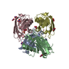
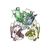
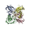
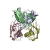
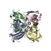
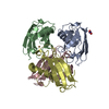
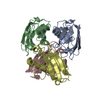
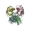
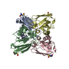
| Title | AZURIN MUTANT M121A-AZIDE | |||||||||
 Components Components | AZURIN AZIDE | |||||||||
 Keywords Keywords | ELECTRON TRANSPORT | |||||||||
| Function / homology |  Function and homology information Function and homology informationtransition metal ion binding / periplasmic space / electron transfer activity / copper ion binding / zinc ion binding / identical protein binding / plasma membrane Similarity search - Function | |||||||||
| Biological species |   Pseudomonas aeruginosa (bacteria) Pseudomonas aeruginosa (bacteria) | |||||||||
| Method |  X-RAY DIFFRACTION / Resolution: 2.3 Å X-RAY DIFFRACTION / Resolution: 2.3 Å | |||||||||
 Authors Authors | Tsai, L.-C. / Bonander, N. / Harata, K. / Karlsson, B.G. / Vanngard, T. / Langer, V. / Sjolin, L. | |||||||||
 Citation Citation | Journal: Acta Crystallogr.,Sect.D / Year: 1996 Title: Mutant Met121Ala of Pseudomonas aeruginosa azurin and its azide derivative: crystal structures and spectral properties. Authors: Tsai, L.C. / Bonander, N. / Harata, K. / Karlsson, G. / Vanngard, T. / Langer, V. / Sjolin, L. | |||||||||
| History |
|
- Structure visualization
| Structure viewer | Molecule: MolmilJmol/JSmol |
|---|
- Downloads & links
-Download
| PDBx/mmCIF format | 2tsb.cif.gz | 111.3 KB | Display | PDBx/mmCIF format |
|---|---|---|---|---|
| PDB format | pdb2tsb.ent.gz | 87.2 KB | Display | PDB format |
| PDBx/mmJSON format | 2tsb.json.gz | Tree view | PDBx/mmJSON format | |
| Others |  Other downloads Other downloads |
-Validation report
| Arichive directory | https://data.pdbj.org/pub/pdb/validation_reports/ts/2tsbftp://data.pdbj.org/pub/pdb/validation_reports/ts/2tsb | HTTPS FTP |
|---|
-Related structure data


-Links
 PDBj
PDBj- Assembly
| Deposited unit | 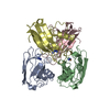
| ||||||||
|---|---|---|---|---|---|---|---|---|---|
| 1 |
| ||||||||
| Unit cell |
|
-Components
| #1: Protein | Mass: 13901.681 Da / Num. of mol.: 4 / Mutation: M121A Source method: isolated from a genetically manipulated source Source: (gene. exp.) Pseudomonas aeruginosa (bacteria) / Plasmid: PUC18/19, M13MP18/19 / Production host: #2: Chemical | ChemComp-AZI /   Mass: 42.020 Da / Num. of mol.: 4 / Source method: obtained synthetically / Formula: N3 Mass: 42.020 Da / Num. of mol.: 4 / Source method: obtained synthetically / Formula: N3#3: Chemical | ChemComp-CU /   Mass: 63.546 Da / Num. of mol.: 4 / Source method: obtained synthetically / Formula: Cu Mass: 63.546 Da / Num. of mol.: 4 / Source method: obtained synthetically / Formula: Cu#4: Water | ChemComp-HOH / |  Mass: 18.015 Da / Num. of mol.: 224 / Source method: isolated from a natural source / Formula: H2O Mass: 18.015 Da / Num. of mol.: 224 / Source method: isolated from a natural source / Formula: H2OHas protein modification | Y | |
|---|
-Experimental details
-Experiment
| Experiment | Method: X-RAY DIFFRACTION / Number of used crystals: 1 |
|---|
- Sample preparation
| Crystal | Density Matthews: 2.34 Å3/Da / Density % sol: 47.45 % | |||||||||||||||
|---|---|---|---|---|---|---|---|---|---|---|---|---|---|---|---|---|
| Crystal grow | *PLUS Temperature: 297-298 K / pH: 5.1 / Method: unknown | |||||||||||||||
| Components of the solutions | *PLUS
|
-Data collection
| Diffraction source | Wavelength: 1.5418 |
|---|---|
| Detector | Type: ENRAF-NONIUS FAST / Detector: DIFFRACTOMETER / Date: Jan 11, 1995 |
| Radiation | Monochromatic (M) / Laue (L): M / Scattering type: x-ray |
| Radiation wavelength | Wavelength: 1.5418 Å / Relative weight: 1 |
| Reflection | Resolution: 2.3→10.8 Å / Num. obs: 18193 / % possible obs: 84 % / Observed criterion σ(I): 0 / Rmerge(I) obs: 0.079 |
| Reflection | *PLUS Num. measured all: 36479 |
- Processing
| Software |
| ||||||||||||||||||||||||||||||||||||||||||||||||||||||||||||
|---|---|---|---|---|---|---|---|---|---|---|---|---|---|---|---|---|---|---|---|---|---|---|---|---|---|---|---|---|---|---|---|---|---|---|---|---|---|---|---|---|---|---|---|---|---|---|---|---|---|---|---|---|---|---|---|---|---|---|---|---|---|
| Refinement | Resolution: 2.3→8 Å / σ(F): 0
| ||||||||||||||||||||||||||||||||||||||||||||||||||||||||||||
| Displacement parameters | Biso mean: 27.17 Å2 | ||||||||||||||||||||||||||||||||||||||||||||||||||||||||||||
| Refine analyze | Luzzati coordinate error obs: 0.22 Å | ||||||||||||||||||||||||||||||||||||||||||||||||||||||||||||
| Refinement step | Cycle: LAST / Resolution: 2.3→8 Å
| ||||||||||||||||||||||||||||||||||||||||||||||||||||||||||||
| Refine LS restraints |
| ||||||||||||||||||||||||||||||||||||||||||||||||||||||||||||
| Software | *PLUS Name: X-PLOR / Classification: refinement | ||||||||||||||||||||||||||||||||||||||||||||||||||||||||||||
| Refinement | *PLUS | ||||||||||||||||||||||||||||||||||||||||||||||||||||||||||||
| Solvent computation | *PLUS | ||||||||||||||||||||||||||||||||||||||||||||||||||||||||||||
| Displacement parameters | *PLUS | ||||||||||||||||||||||||||||||||||||||||||||||||||||||||||||
| Refine LS restraints | *PLUS
|
