 Movie
Movie Controller


+ Open data

- Basic information
| Entry | Database: PDB / ID: 1cc3 | ||||||
|---|---|---|---|---|---|---|---|
| Title | PURPLE CUA CENTER | ||||||
 Components Components | PROTEIN (CUA AZURIN) | ||||||
 Keywords Keywords | ELECTRON TRANSPORT / COPPER-A | ||||||
| Function / homology |  Function and homology information Function and homology informationtransition metal ion binding / electron transfer activity / periplasmic space / copper ion binding / zinc ion binding / identical protein binding / plasma membrane Similarity search - Function | ||||||
| Biological species |   Pseudomonas aeruginosa (bacteria) Pseudomonas aeruginosa (bacteria) | ||||||
| Method |  X-RAY DIFFRACTION / MOLECULAR REPLACEMENT / Resolution: 1.65 Å X-RAY DIFFRACTION / MOLECULAR REPLACEMENT / Resolution: 1.65 Å | ||||||
 Authors Authors | Robinson, H. / Ang, M.C. / Gao, Y.-G. / Hay, M.T. / Lu, Y. / Wang, A.H.-J. | ||||||
 Citation Citation | Journal: Biochemistry / Year: 1999 Title: Structural basis of electron transfer modulation in the purple CuA center. Authors: Robinson, H. / Ang, M.C. / Gao, Y.G. / Hay, M.T. / Lu, Y. / Wang, A.H. | ||||||
| History |
|
- Structure visualization
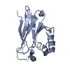
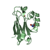
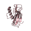
| Structure viewer | Molecule: MolmilJmol/JSmol |
|---|
- Downloads & links
-Download
| PDBx/mmCIF format | 1cc3.cif.gz | 75.3 KB | Display | PDBx/mmCIF format |
|---|---|---|---|---|
| PDB format | pdb1cc3.ent.gz | 54.2 KB | Display | PDB format |
| PDBx/mmJSON format | 1cc3.json.gz | Tree view | PDBx/mmJSON format | |
| Others |  Other downloads Other downloads |
-Validation report
| Arichive directory | https://data.pdbj.org/pub/pdb/validation_reports/cc/1cc3ftp://data.pdbj.org/pub/pdb/validation_reports/cc/1cc3 | HTTPS FTP |
|---|
-Related structure data
| Related structure data |  4azuS S: Starting model for refinement |
|---|---|
| Similar structure data |


-Links
 PDBj
PDBj- Assembly
| Deposited unit | 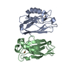
| ||||||||
|---|---|---|---|---|---|---|---|---|---|
| 1 | 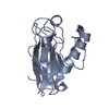
| ||||||||
| 2 | 
| ||||||||
| Unit cell |
|
-Components
| #1: Protein | Mass: 14189.080 Da / Num. of mol.: 2 Source method: isolated from a genetically manipulated source Details: AZURINE WITH THE FOLLOWING MUTATIONS: THE LOOP 113-118 TFPGHS WAS REPLACED WITH 113-120 SELCGINH Source: (gene. exp.) Pseudomonas aeruginosa (bacteria) / Description: LOOP DIRECTED MUTAGENESIS / Production host: #2: Chemical | ChemComp-CU /   Mass: 63.546 Da / Num. of mol.: 4 / Source method: obtained synthetically / Formula: Cu Mass: 63.546 Da / Num. of mol.: 4 / Source method: obtained synthetically / Formula: Cu#3: Water | ChemComp-HOH / |  Mass: 18.015 Da / Num. of mol.: 506 / Source method: isolated from a natural source / Formula: H2O Mass: 18.015 Da / Num. of mol.: 506 / Source method: isolated from a natural source / Formula: H2OHas protein modification | Y | |
|---|
-Experimental details
-Experiment
| Experiment | Method: X-RAY DIFFRACTION / Number of used crystals: 1 |
|---|
- Sample preparation
| Crystal | Density Matthews: 1.96 Å3/Da / Density % sol: 37 % | ||||||||||||||||||||||||||||||||||||||||||||||||
|---|---|---|---|---|---|---|---|---|---|---|---|---|---|---|---|---|---|---|---|---|---|---|---|---|---|---|---|---|---|---|---|---|---|---|---|---|---|---|---|---|---|---|---|---|---|---|---|---|---|
| Crystal grow | pH: 5.1 / Details: pH 5.1 | ||||||||||||||||||||||||||||||||||||||||||||||||
| Crystal | *PLUS | ||||||||||||||||||||||||||||||||||||||||||||||||
| Crystal grow | *PLUS Temperature: 5 ℃ / Method: vapor diffusion | ||||||||||||||||||||||||||||||||||||||||||||||||
| Components of the solutions | *PLUS
|
-Data collection
| Diffraction | Mean temperature: 123 K |
|---|---|
| Diffraction source | Source: ROTATING ANODE / Type: RIGAKU RU200 / Wavelength: 1.5418 |
| Detector | Type: RIGAKU / Detector: IMAGE PLATE / Date: Sep 11, 1998 |
| Radiation | Monochromator: GRAPHITE / Protocol: SINGLE WAVELENGTH / Monochromatic (M) / Laue (L): M / Scattering type: x-ray |
| Radiation wavelength | Wavelength: 1.5418 Å / Relative weight: 1 |
| Reflection | Resolution: 1.65→20 Å / Num. obs: 26467 / % possible obs: 94.1 % / Observed criterion σ(I): 0 / Redundancy: 3 % / Rmerge(I) obs: 0.105 / Net I/σ(I): 17 |
| Reflection shell | Resolution: 1.65→1.75 Å / Redundancy: 2.6 % / Rmerge(I) obs: 0.76 / Mean I/σ(I) obs: 3.3 / % possible all: 96.4 |
| Reflection | *PLUS % possible obs: 97.8 % |
- Processing
| Software |
| |||||||||||||||||||||||||||||||||
|---|---|---|---|---|---|---|---|---|---|---|---|---|---|---|---|---|---|---|---|---|---|---|---|---|---|---|---|---|---|---|---|---|---|---|
| Refinement | Method to determine structure: MOLECULAR REPLACEMENT Starting model: 4AZU Resolution: 1.65→20 Å / Num. parameters: 9959 / Num. restraintsaints: 8058 / Cross valid method: THROUGHOUT UNTIL END / σ(F): 0 / Stereochemistry target values: ENGH AND HUBER / Details: NO RESTRAINTS APPLIED TO COPPER ATOMS
| |||||||||||||||||||||||||||||||||
| Solvent computation | Solvent model: MOEWS & KRETSINGER | |||||||||||||||||||||||||||||||||
| Refine analyze | Num. disordered residues: 0 / Occupancy sum hydrogen: 0 / Occupancy sum non hydrogen: 2486 | |||||||||||||||||||||||||||||||||
| Refinement step | Cycle: LAST / Resolution: 1.65→20 Å
| |||||||||||||||||||||||||||||||||
| Refine LS restraints |
| |||||||||||||||||||||||||||||||||
| Software | *PLUS Name: SHELXL-97 / Classification: refinement | |||||||||||||||||||||||||||||||||
| Refinement | *PLUS Lowest resolution: 20 Å / σ(F): 0 / % reflection Rfree: 5 % / Rfactor obs: 0.189 / Rfactor Rwork: 0.2 | |||||||||||||||||||||||||||||||||
| Solvent computation | *PLUS | |||||||||||||||||||||||||||||||||
| Displacement parameters | *PLUS |
