 Movie
Movie Controller


+ Open data

- Basic information
| Entry | Database: PDB / ID: 1jvo | ||||||
|---|---|---|---|---|---|---|---|
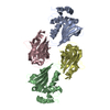
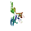
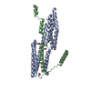
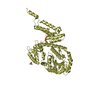
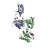
| Title | Azurin dimer, crosslinked via disulfide bridge | ||||||
 Components Components | Azurin | ||||||
 Keywords Keywords | ELECTRON TRANSPORT / cupredoxin / electron transfer / covalent crosslink | ||||||
| Function / homology |  Function and homology information Function and homology informationtransition metal ion binding / periplasmic space / electron transfer activity / copper ion binding / zinc ion binding / identical protein binding / plasma membrane Similarity search - Function | ||||||
| Biological species |   Pseudomonas aeruginosa (bacteria) Pseudomonas aeruginosa (bacteria) | ||||||
| Method |  X-RAY DIFFRACTION / SYNCHROTRON / MOLECULAR REPLACEMENT / Resolution: 2.75 Å X-RAY DIFFRACTION / SYNCHROTRON / MOLECULAR REPLACEMENT / Resolution: 2.75 Å | ||||||
 Authors Authors | van Amsterdam, I.M.C. / Ubbink, M. / Einsle, O. / Messerschmidt, A. / Merli, A. / Cavazzini, D. / Rossi, G.L. / Canters, G.W. | ||||||
 Citation Citation | Journal: Nat.Struct.Biol. / Year: 2002 Title: Dramatic modulation of electron transfer in protein complexes by crosslinking Authors: van Amsterdam, I.M.C. / Ubbink, M. / Einsle, O. / Messerschmidt, A. / Merli, A. / Cavazzini, D. / Rossi, G.L. / Canters, G.W. #1: Journal: Chemistry / Year: 2001Title: Effects of Dimerization on Protein Electron Transfer Authors: van Amsterdam, I.M.C. / Ubbink, M. / Jeuken, L.J.C. / Verbeet, M.P. / Einsle, O. / Messerschmidt, A. / Canters, G.W. | ||||||
| History |
|
- Structure visualization
| Structure viewer | Molecule: MolmilJmol/JSmol |
|---|
- Downloads & links
-Download
| PDBx/mmCIF format | 1jvo.cif.gz | 287.3 KB | Display | PDBx/mmCIF format |
|---|---|---|---|---|
| PDB format | pdb1jvo.ent.gz | 237.5 KB | Display | PDB format |
| PDBx/mmJSON format | 1jvo.json.gz | Tree view | PDBx/mmJSON format | |
| Others |  Other downloads Other downloads |
-Validation report
| Arichive directory | https://data.pdbj.org/pub/pdb/validation_reports/jv/1jvoftp://data.pdbj.org/pub/pdb/validation_reports/jv/1jvo | HTTPS FTP |
|---|
-Related structure data


-Links
 PDBj
PDBj- Assembly
| Deposited unit | 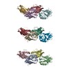
| ||||||||||
|---|---|---|---|---|---|---|---|---|---|---|---|
| 1 | 
| ||||||||||
| 2 | 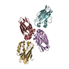
| ||||||||||
| 3 | 
| ||||||||||
| Unit cell |
| ||||||||||
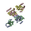
| Details | FOUR MONOMERS GENERATE A TETRAMER ACTIVE IN INTERMOLECULAR ELECTRON TRANSFER. THE ASYMMETRIC UNIT CONTAINS THREE SUCH TETRAMERS. PROGRAM PISA DOES NOT PREDICT THOSE ASSEMBLIES BECAUSE THE INDIVIDUAL INTERMOLECULAR DISULFIDE BOND DOES NOT CREATE SUFFICIENT BURIED AREA. |
-Components
| #1: Protein | Mass: 13950.839 Da / Num. of mol.: 12 / Fragment: Azurin / Mutation: N42C Source method: isolated from a genetically manipulated source Source: (gene. exp.) Pseudomonas aeruginosa (bacteria) / Gene: azu / Plasmid: pGK22 / Production host: #2: Chemical | ChemComp-CU /   Mass: 63.546 Da / Num. of mol.: 12 / Source method: obtained synthetically / Formula: Cu Mass: 63.546 Da / Num. of mol.: 12 / Source method: obtained synthetically / Formula: CuHas protein modification | Y | |
|---|
-Experimental details
-Experiment
| Experiment | Method: X-RAY DIFFRACTION / Number of used crystals: 1 |
|---|
- Sample preparation
| Crystal | Density Matthews: 2.35 Å3/Da / Density % sol: 47.73 % | ||||||||||||||||||||||||
|---|---|---|---|---|---|---|---|---|---|---|---|---|---|---|---|---|---|---|---|---|---|---|---|---|---|
| Crystal grow | Temperature: 293 K / Method: vapor diffusion, sitting drop / pH: 8.5 Details: PEG 8000, Tris/HCl, pH 8.5, VAPOR DIFFUSION, SITTING DROP at 293K, temperature 293.0K | ||||||||||||||||||||||||
| Crystal grow | *PLUS | ||||||||||||||||||||||||
| Components of the solutions | *PLUS
|
-Data collection
| Diffraction | Mean temperature: 100 K |
|---|---|
| Diffraction source | Source: SYNCHROTRON / Site: MPG/DESY, HAMBURG  / Beamline: BW6 / Wavelength: 1.05 Å / Beamline: BW6 / Wavelength: 1.05 Å |
| Detector | Type: MARRESEARCH / Detector: CCD / Date: Oct 9, 2000 |
| Radiation | Monochromator: GRAPHITE / Protocol: SINGLE WAVELENGTH / Monochromatic (M) / Laue (L): M / Scattering type: x-ray |
| Radiation wavelength | Wavelength: 1.05 Å / Relative weight: 1 |
| Reflection | Resolution: 2.75→50 Å / Num. all: 41247 / Num. obs: 35555 / % possible obs: 86.2 % / Observed criterion σ(I): 3 / Redundancy: 1.9 % / Biso Wilson estimate: 60.1 Å2 / Rsym value: 0.101 / Net I/σ(I): 7.1 |
| Reflection shell | Resolution: 2.75→2.85 Å / Redundancy: 1.4 % / Mean I/σ(I) obs: 1.6 / Num. unique all: 3588 / Rsym value: 0.404 / % possible all: 88.2 |
| Reflection | *PLUS Num. obs: 42605 / Rmerge(I) obs: 0.108 |
| Reflection shell | *PLUS % possible obs: 86.4 % / Rmerge(I) obs: 0.484 |
- Processing
| Software |
| |||||||||||||||||||||||||
|---|---|---|---|---|---|---|---|---|---|---|---|---|---|---|---|---|---|---|---|---|---|---|---|---|---|---|
| Refinement | Method to determine structure: MOLECULAR REPLACEMENT / Resolution: 2.75→50 Å / σ(F): 0 / Stereochemistry target values: Engh & Huber / Details: CHAINS F AND K ARE PARTIALLY DISORDERED
| |||||||||||||||||||||||||
| Displacement parameters |
| |||||||||||||||||||||||||
| Refinement step | Cycle: LAST / Resolution: 2.75→50 Å
| |||||||||||||||||||||||||
| Refine LS restraints |
| |||||||||||||||||||||||||
| Software | *PLUS Name: CNS / Version: 1 / Classification: refinement | |||||||||||||||||||||||||
| Refinement | *PLUS Lowest resolution: 50 Å / σ(F): 0 / % reflection Rfree: 3 % / Rfactor obs: 0.239 / Rfactor Rfree: 0.293 | |||||||||||||||||||||||||
| Solvent computation | *PLUS | |||||||||||||||||||||||||
| Displacement parameters | *PLUS | |||||||||||||||||||||||||
| Refine LS restraints | *PLUS Type: c_angle_deg / Dev ideal: 1.34 |
