 Movie
Movie Controller

[English] 日本語
 Yorodumi
- PDB-1dt9: THE CRYSTAL STRUCTURE OF HUMAN EUKARYOTIC RELEASE FACTOR ERF1-MEC... -
+ Open data

- Basic information
| Entry | Database: PDB / ID: 1dt9 | ||||||
|---|---|---|---|---|---|---|---|
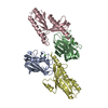
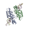
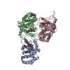
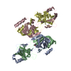
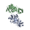
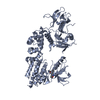
| Title | THE CRYSTAL STRUCTURE OF HUMAN EUKARYOTIC RELEASE FACTOR ERF1-MECHANISM OF STOP CODON RECOGNITION AND PEPTIDYL-TRNA HYDROLYSIS | ||||||
 Components Components | PROTEIN (EUKARYOTIC PEPTIDE CHAIN RELEASE FACTOR SUBUNIT 1) | ||||||
 Keywords Keywords | TRANSLATION / erf1 / trna mimicry / protein sythesis / stop codon recognition / peptidyl-trna hydrolysis | ||||||
| Function / homology |  Function and homology information Function and homology informationtranslation termination factor activity / translation release factor complex / cytoplasmic translational termination / regulation of translational termination / translation release factor activity / translation release factor activity, codon specific / protein methylation / sequence-specific mRNA binding / peptidyl-tRNA hydrolase activity / nuclear-transcribed mRNA catabolic process, nonsense-mediated decay ...translation termination factor activity / translation release factor complex / cytoplasmic translational termination / regulation of translational termination / translation release factor activity / translation release factor activity, codon specific / protein methylation / sequence-specific mRNA binding / peptidyl-tRNA hydrolase activity / nuclear-transcribed mRNA catabolic process, nonsense-mediated decay / Protein hydroxylation / Eukaryotic Translation Termination / Nonsense Mediated Decay (NMD) independent of the Exon Junction Complex (EJC) / translational termination / Nonsense Mediated Decay (NMD) enhanced by the Exon Junction Complex (EJC) / cytosolic ribosome / Regulation of expression of SLITs and ROBOs / ribosome binding / RNA binding / cytosol / cytoplasm Similarity search - Function | ||||||
| Biological species |  Homo sapiens (human) Homo sapiens (human) | ||||||
| Method |  X-RAY DIFFRACTION / SYNCHROTRON / Resolution: 2.7 Å X-RAY DIFFRACTION / SYNCHROTRON / Resolution: 2.7 Å | ||||||
 Authors Authors | Frolova, L. | ||||||
 Citation Citation | Journal: Cell(Cambridge,Mass.) / Year: 2000 Title: The crystal structure of human eukaryotic release factor eRF1--mechanism of stop codon recognition and peptidyl-tRNA hydrolysis. Authors: Song, H. / Mugnier, P. / Das, A.K. / Webb, H.M. / Evans, D.R. / Tuite, M.F. / Hemmings, B.A. / Barford, D. #1: Journal: RNA / Year: 1999Title: Mutations in the Highly Conserved GGQ Motif of Class 1 Polypeptide Release Factors Abolish Ability of Human eRF1 to Trigger Peptidyl-tRNA Hydrolysis. Authors: Frolova, L.Y. / Tsivkovskii, R.Y. / Sivolobova, G.F. / Oparina, N.Y. / Serpinsky, O.I. / Blinov, V.M. / Tatkov, S.I. / Kisselev, L.L. #2: Journal: Nature / Year: 1994Title: A Highly Conserved Eukaryotic Protein Family Possesing Properties of Polypeptide Chain Release Factor Authors: Frolova, L. / Le Goff, X. / Rasmussen, H.H. / Cheperegin, S. / Drugeon, G. / Kress, M. / Arman, I. / Haenni, A.L. / Celis, J.E. / Philippe, M. / Justesen, J. / Kisselev, L. | ||||||
| History |
|
- Structure visualization
| Structure viewer | Molecule: MolmilJmol/JSmol |
|---|
- Downloads & links
-Download
| PDBx/mmCIF format | 1dt9.cif.gz | 92.1 KB | Display | PDBx/mmCIF format |
|---|---|---|---|---|
| PDB format | pdb1dt9.ent.gz | 71.3 KB | Display | PDB format |
| PDBx/mmJSON format | 1dt9.json.gz | Tree view | PDBx/mmJSON format | |
| Others |  Other downloads Other downloads |
-Validation report
| Arichive directory | https://data.pdbj.org/pub/pdb/validation_reports/dt/1dt9ftp://data.pdbj.org/pub/pdb/validation_reports/dt/1dt9 | HTTPS FTP |
|---|
-Related structure data
| Similar structure data |
|---|


-Links
 PDBj
PDBj


- Assembly
| Deposited unit | 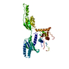
| ||||||||
|---|---|---|---|---|---|---|---|---|---|
| 1 |
| ||||||||
| Unit cell |
|
-Components
| #1: Protein | Mass: 49092.742 Da / Num. of mol.: 1 / Source method: isolated from a natural source / Source: (natural) Homo sapiens (human) / References: UniProt: P62495 |
|---|---|
| #2: Water | ChemComp-HOH /  Mass: 18.015 Da / Num. of mol.: 110 / Source method: isolated from a natural source / Formula: H2O Mass: 18.015 Da / Num. of mol.: 110 / Source method: isolated from a natural source / Formula: H2O |
-Experimental details
-Experiment
| Experiment | Method: X-RAY DIFFRACTION / Number of used crystals: 1 |
|---|
- Sample preparation
| Crystal | Density Matthews: 2.94 Å3/Da / Density % sol: 58.17 % | ||||||||||||||||||||||||||||||||||||
|---|---|---|---|---|---|---|---|---|---|---|---|---|---|---|---|---|---|---|---|---|---|---|---|---|---|---|---|---|---|---|---|---|---|---|---|---|---|
| Crystal grow | Temperature: 293 K / Method: vapor diffusion, hanging drop / pH: 7.5 Details: Hepes, PEG4000, Glycerol, NaCl, pH 7.5, VAPOR DIFFUSION, HANGING DROP, temperature 293K | ||||||||||||||||||||||||||||||||||||
| Crystal grow | *PLUS Temperature: 20 ℃Details: protein was mixed with equal volume of reservoir solution | ||||||||||||||||||||||||||||||||||||
| Components of the solutions | *PLUS
|
-Data collection
| Diffraction source | Source: SYNCHROTRON / Site: EMBL/DESY, HAMBURG  / Beamline: BW7A / Beamline: BW7A |
|---|---|
| Detector | Detector: CCD |
| Radiation | Protocol: SINGLE WAVELENGTH / Monochromatic (M) / Laue (L): M / Scattering type: x-ray |
| Radiation wavelength | Relative weight: 1 |
| Reflection | Resolution: 2.7→25 Å / Num. obs: 82505 / % possible obs: 99.2 % / Observed criterion σ(F): 0 / Observed criterion σ(I): 0 / Redundancy: 4.9 % / Rmerge(I) obs: 0.05 / Net I/σ(I): 8.3 |
| Reflection shell | Resolution: 2.7→25 Å / Redundancy: 3.5 % / Rmerge(I) obs: 0.406 / Num. unique all: 16712 / % possible all: 98 |
| Reflection | *PLUS Num. obs: 16712 / Num. measured all: 82505 |
| Reflection shell | *PLUS % possible obs: 98 % / Mean I/σ(I) obs: 1.9 |
- Processing
| Software |
| ||||||||||||||||
|---|---|---|---|---|---|---|---|---|---|---|---|---|---|---|---|---|---|
| Refinement | Resolution: 2.7→20 Å / σ(F): 0 / σ(I): 0
| ||||||||||||||||
| Refinement step | Cycle: LAST / Resolution: 2.7→20 Å
| ||||||||||||||||
| Refine LS restraints |
|
