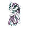
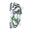
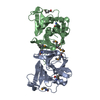
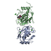
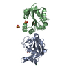
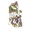
The biological unit is a dimer. There are 2 such biological units (dimers) present in the asymmetric unit (One dimer is formed by chains A & B and the other by chains C & D).
-
Components
#1: Protein
Sortase
Mass: 21109.846 Da / Num. of mol.: 3 / Fragment: CATALYTIC DOMAIN, UNP residues 82-247 / Mutation: T176A Source method: isolated from a genetically manipulated source Source: (gene. exp.) Streptococcus pneumoniae (bacteria) / Strain: ATCC BAA-255 / R6 / Gene: sortase A, spr1098, srtA / Plasmid: pET28c / Production host: Escherichia coli (E. coli) / Strain (production host): BL21 (DE3) / References: UniProt: Q8DPM3, sortase A
#2: Protein
Sortase
Mass: 21125.846 Da / Num. of mol.: 1 / Fragment: CATALYTIC DOMAIN, UNP residues 82-247 / Mutation: T176A Source method: isolated from a genetically manipulated source Source: (gene. exp.) Streptococcus pneumoniae (bacteria) / Strain: ATCC BAA-255 / R6 / Gene: sortase A, spr1098, srtA / Plasmid: pET28c / Production host: Escherichia coli (E. coli) / Strain (production host): BL21 (DE3) / References: UniProt: Q8DPM3, sortase A
Mass: 18.015 Da / Num. of mol.: 54 / Source method: isolated from a natural source / Formula: H2O
Has protein modification
Y
-
Experimental details
-
Experiment
Experiment
Method: X-RAY DIFFRACTION / Number of used crystals: 1
-
Sample preparation
Crystal
Density Matthews: 2.77 Å3/Da / Density % sol: 55.55 % / Mosaicity: 1.52 °
Crystal grow
Temperature: 293 K / Method: microbatch, under oil / pH: 7 Details: 0.2 M tri-ammonium citrate, pH 7.0, 20% (w/v) PEG 3350, 40% (v/v) 1,3-Butanediol, Microbatch, Under oil, temperature 293K
-
Data collection
Diffraction
Mean temperature: 100 K
Diffraction source
Source: ROTATING ANODE / Type: OTHER / Wavelength: 1.5418 Å
Detector
Type: MAR scanner 345 mm plate / Detector: IMAGE PLATE / Date: Oct 8, 2011 / Details: MIRRORS
Radiation
Monochromator: OSMIC MIRROR / Protocol: SINGLE WAVELENGTH / Monochromatic (M) / Laue (L): M / Scattering type: x-ray
Radiation wavelength
Wavelength: 1.5418 Å / Relative weight: 1
Reflection
Resolution: 2.7→67.492 Å / Num. all: 25168 / Num. obs: 25168 / % possible obs: 99.2 % / Redundancy: 7.5 % / Rsym value: 0.094 / Net I/σ(I): 14.3
Reflection shell
Diffraction-ID: 1
Resolution (Å)
Redundancy (%)
Rmerge(I) obs
Mean I/σ(I) obs
Num. measured all
Num. unique all
Rpim(I) all
Rsym value
Net I/σ(I) obs
% possible all
2.7-2.85
7.4
0.529
1.5
26715
3621
0.206
0.529
3.8
98.6
2.85-3.02
7.5
0.368
2.1
25840
3468
0.142
0.368
5.3
98.8
3.02-3.23
7.5
0.222
3.5
24210
3225
0.086
0.222
8.3
99
3.23-3.49
7.4
0.151
4.2
22594
3039
0.059
0.151
11.6
99.3
3.49-3.82
7.5
0.096
7.2
20874
2801
0.038
0.096
16.9
99.5
3.82-4.27
7.5
0.075
7.8
19137
2559
0.029
0.075
20
99.7
4.27-4.93
7.5
0.06
10.1
16786
2231
0.024
0.06
24.8
99.7
4.93-6.04
7.5
0.066
9.1
14436
1915
0.026
0.066
24.1
99.9
6.04-8.54
7.5
0.056
9.5
11162
1497
0.022
0.056
26.5
100
8.54-31.841
7.3
0.052
9.5
5905
812
0.021
0.052
31.3
98
-
Processing
Software
Name
Version
Classification
NB
SCALA
3.3.16
datascaling
REFMAC
5.7.0029
refinement
PDB_EXTRACT
3.14
dataextraction
MAR345dtb
datacollection
MOSFLM
datareduction
PHASER
phasing
Refinement
Method to determine structure: MOLECULAR REPLACEMENT / Resolution: 2.7→30.71 Å / Cor.coef. Fo:Fc: 0.951 / Cor.coef. Fo:Fc free: 0.926 / WRfactor Rfree: 0.2245 / WRfactor Rwork: 0.1727 / Occupancy max: 1 / Occupancy min: 0.5 / FOM work R set: 0.8449 / SU B: 23.863 / SU ML: 0.228 / SU R Cruickshank DPI: 0.6869 / SU Rfree: 0.3028 / Cross valid method: THROUGHOUT / σ(F): 0 / ESU R: 0.687 / ESU R Free: 0.303 / Stereochemistry target values: MAXIMUM LIKELIHOOD Details: HYDROGENS HAVE BEEN ADDED IN THE RIDING POSITIONS U VALUES : WITH TLS ADDED. Oxidized CYS (CSO) was modeled in place of CYS 207 in Chain B.
Rfactor
Num. reflection
% reflection
Selection details
Rfree
0.2339
1273
5.1 %
RANDOM
Rwork
0.181
-
-
-
obs
0.1836
25152
99.19 %
-
Solvent computation
Ion probe radii: 0.8 Å / Shrinkage radii: 0.8 Å / VDW probe radii: 1.2 Å / Solvent model: MASK
In the structure databanks used in Yorodumi, some data are registered as the other names, "COVID-19 virus" and "2019-nCoV". Here are the details of the virus and the list of structure data.
Jan 31, 2019. EMDB accession codes are about to change! (news from PDBe EMDB page)
EMDB accession codes are about to change! (news from PDBe EMDB page)
The allocation of 4 digits for EMDB accession codes will soon come to an end. Whilst these codes will remain in use, new EMDB accession codes will include an additional digit and will expand incrementally as the available range of codes is exhausted. The current 4-digit format prefixed with “EMD-” (i.e. EMD-XXXX) will advance to a 5-digit format (i.e. EMD-XXXXX), and so on. It is currently estimated that the 4-digit codes will be depleted around Spring 2019, at which point the 5-digit format will come into force.
The EM Navigator/Yorodumi systems omit the EMD- prefix.
Related info.:Q: What is EMD? / ID/Accession-code notation in Yorodumi/EM Navigator
Yorodumi is a browser for structure data from EMDB, PDB, SASBDB, etc.
This page is also the successor to EM Navigator detail page, and also detail information page/front-end page for Omokage search.
The word "yorodu" (or yorozu) is an old Japanese word meaning "ten thousand". "mi" (miru) is to see.
Related info.:EMDB / PDB / SASBDB / Comparison of 3 databanks / Yorodumi Search / Aug 31, 2016. New EM Navigator & Yorodumi / Yorodumi Papers / Jmol/JSmol / Function and homology information / Changes in new EM Navigator and Yorodumi
 Movie
Movie Controller


 Open data
 Basic information
 Components
 Keywords
 Function and homology information
 Streptococcus pneumoniae (bacteria)
 X-RAY DIFFRACTION /
  Authors
 Citation
 Structure visualization
 Downloads & links
 Other downloads


 PDBj
PDBj Assembly
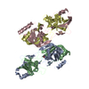

 Mass: 18.015 Da / Num. of mol.: 54 / Source method: isolated from a natural source / Formula: H2O
 Sample preparation
 Processing