 Movie
Movie Controller

[English] 日本語
 Yorodumi
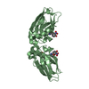
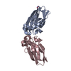
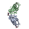
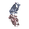
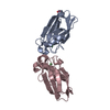
- PDB-1jvl: Azurin dimer, covalently crosslinked through bis-maleimidomethylether -
+ Open data

- Basic information
| Entry | Database: PDB / ID: 1jvl | ||||||
|---|---|---|---|---|---|---|---|
| Title | Azurin dimer, covalently crosslinked through bis-maleimidomethylether | ||||||
 Components Components | Azurin | ||||||
 Keywords Keywords | ELECTRON TRANSPORT / cupredoxin / electron transfer / covalent crosslink | ||||||
| Function / homology |  Function and homology information Function and homology informationtransition metal ion binding / electron transfer activity / periplasmic space / copper ion binding / zinc ion binding / identical protein binding / plasma membrane Similarity search - Function | ||||||
| Biological species |   Pseudomonas aeruginosa (bacteria) Pseudomonas aeruginosa (bacteria) | ||||||
| Method |  X-RAY DIFFRACTION / SYNCHROTRON / MOLECULAR REPLACEMENT / Resolution: 2 Å X-RAY DIFFRACTION / SYNCHROTRON / MOLECULAR REPLACEMENT / Resolution: 2 Å | ||||||
 Authors Authors | van Amsterdam, I.M.C. / Ubbink, M. / Einsle, O. / Messerschmidt, A. / Merli, A. / Cavazzini, D. / Rossi, G.L. / Canters, G.W. | ||||||
 Citation Citation | Journal: Nat.Struct.Biol. / Year: 2002 Title: Dramatic modulation of electron transfer in protein complexes by crosslinking Authors: van Amsterdam, I.M.C. / Ubbink, M. / Einsle, O. / Messerschmidt, A. / Merli, A. / Cavazzini, D. / Rossi, G.L. / Canters, G.W. #1: Journal: Chemistry / Year: 2001Title: Effects of Dimerization on Protein Electron Transfer Authors: van Amsterdam, I.M.C. / Ubbink, M. / Jeuken, L.J.C. / Verbeet, M.P. / Einsle, O. / Messerschmidt, A. / Canters, G.W. | ||||||
| History |
|
- Structure visualization
| Structure viewer | Molecule: MolmilJmol/JSmol |
|---|
- Downloads & links
-Download
| PDBx/mmCIF format | 1jvl.cif.gz | 65.8 KB | Display | PDBx/mmCIF format |
|---|---|---|---|---|
| PDB format | pdb1jvl.ent.gz | 47.9 KB | Display | PDB format |
| PDBx/mmJSON format | 1jvl.json.gz | Tree view | PDBx/mmJSON format | |
| Others |  Other downloads Other downloads |
-Validation report
| Arichive directory | https://data.pdbj.org/pub/pdb/validation_reports/jv/1jvlftp://data.pdbj.org/pub/pdb/validation_reports/jv/1jvl | HTTPS FTP |
|---|
-Related structure data


-Links
 PDBj
PDBj- Assembly
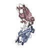
| Deposited unit | 
| ||||||||||
|---|---|---|---|---|---|---|---|---|---|---|---|
| 1 |
| ||||||||||
| Unit cell |
| ||||||||||
| Details | The true spacegroup of the data is P6122, but the structure was refined in P61 to be able to build a dimer, which is linked via BMME across a crystallographic twofold. Strict NCS restraints were applied. |
-Components
-Protein , 1 types, 2 molecules AB
| #1: Protein | Mass: 13950.839 Da / Num. of mol.: 2 / Fragment: Azurin / Mutation: N42C Source method: isolated from a genetically manipulated source Source: (gene. exp.) Pseudomonas aeruginosa (bacteria) / Gene: azu / Plasmid: pGK22 / Production host: |
|---|
-Non-polymers , 5 types, 121 molecules 


| #2: Chemical |  Mass: 63.546 Da / Num. of mol.: 2 / Source method: obtained synthetically / Formula: Cu Mass: 63.546 Da / Num. of mol.: 2 / Source method: obtained synthetically / Formula: Cu#3: Chemical |  Mass: 58.693 Da / Num. of mol.: 2 / Source method: obtained synthetically / Formula: Ni Mass: 58.693 Da / Num. of mol.: 2 / Source method: obtained synthetically / Formula: Ni#4: Chemical |  Mass: 122.143 Da / Num. of mol.: 2 / Source method: obtained synthetically / Formula: C4H12NO3 Mass: 122.143 Da / Num. of mol.: 2 / Source method: obtained synthetically / Formula: C4H12NO3#5: Chemical | ChemComp-OPP / |  Mass: 236.181 Da / Num. of mol.: 1 / Source method: obtained synthetically / Formula: C10H8N2O5 Mass: 236.181 Da / Num. of mol.: 1 / Source method: obtained synthetically / Formula: C10H8N2O5#6: Water | ChemComp-HOH / | Mass: 18.015 Da / Num. of mol.: 114 / Source method: isolated from a natural source / Formula: H2O |
|---|
-Details
| Has protein modification | Y |
|---|
-Experimental details
-Experiment
| Experiment | Method: X-RAY DIFFRACTION / Number of used crystals: 1 |
|---|
- Sample preparation
| Crystal | Density Matthews: 3.48 Å3/Da / Density % sol: 64.65 % | |||||||||||||||||||||||||||||||||||
|---|---|---|---|---|---|---|---|---|---|---|---|---|---|---|---|---|---|---|---|---|---|---|---|---|---|---|---|---|---|---|---|---|---|---|---|---|
| Crystal grow | Temperature: 293 K / Method: vapor diffusion, sitting drop / pH: 8.5 Details: PEG 2000 MME, nickel chloride, Tris/HCl, pH 8.5, VAPOR DIFFUSION, SITTING DROP at 293K | |||||||||||||||||||||||||||||||||||
| Crystal grow | *PLUS pH: 9 | |||||||||||||||||||||||||||||||||||
| Components of the solutions | *PLUS
|
-Data collection
| Diffraction | Mean temperature: 100 K |
|---|---|
| Diffraction source | Source: SYNCHROTRON / Site: MPG/DESY, HAMBURG  / Beamline: BW6 / Wavelength: 1.05 Å / Beamline: BW6 / Wavelength: 1.05 Å |
| Detector | Type: MARRESEARCH / Detector: CCD / Date: Oct 9, 2000 |
| Radiation | Monochromator: GRAPHITE / Protocol: SINGLE WAVELENGTH / Monochromatic (M) / Laue (L): M / Scattering type: x-ray |
| Radiation wavelength | Wavelength: 1.05 Å / Relative weight: 1 |
| Reflection | Resolution: 2→50 Å / Num. all: 25663 / Num. obs: 25535 / % possible obs: 99.5 % / Observed criterion σ(I): 3 / Redundancy: 3.2 % / Biso Wilson estimate: 39.73 Å2 / Rsym value: 0.072 / Net I/σ(I): 15 |
| Reflection shell | Resolution: 2→2.06 Å / Redundancy: 3.1 % / Mean I/σ(I) obs: 2.1 / Num. unique all: 2093 / Rsym value: 0.44 / % possible all: 99.9 |
| Reflection | *PLUS Lowest resolution: 50 Å / Rmerge(I) obs: 0.07 |
| Reflection shell | *PLUS % possible obs: 99.9 % / Rmerge(I) obs: 0.44 |
- Processing
| Software |
| |||||||||||||||||||||||||
|---|---|---|---|---|---|---|---|---|---|---|---|---|---|---|---|---|---|---|---|---|---|---|---|---|---|---|
| Refinement | Method to determine structure: MOLECULAR REPLACEMENT / Resolution: 2→50 Å / Cross valid method: THROUGHOUT / σ(F): 0 / Stereochemistry target values: Engh & Huber
| |||||||||||||||||||||||||
| Displacement parameters |
| |||||||||||||||||||||||||
| Refinement step | Cycle: LAST / Resolution: 2→50 Å
| |||||||||||||||||||||||||
| Refine LS restraints |
| |||||||||||||||||||||||||
| Software | *PLUS Name: CNS / Version: 1 / Classification: refinement | |||||||||||||||||||||||||
| Refinement | *PLUS Highest resolution: 2 Å / Lowest resolution: 50 Å / σ(F): 0 / % reflection Rfree: 4.8 % / Rfactor Rwork: 0.19 | |||||||||||||||||||||||||
| Solvent computation | *PLUS | |||||||||||||||||||||||||
| Displacement parameters | *PLUS | |||||||||||||||||||||||||
| Refine LS restraints | *PLUS Type: c_bond_d / Dev ideal: 0.0054 |
