[English] 日本語
Yorodumi
- PDB-4k0d: Periplasmic sensor domain of sensor histidine kinase, Adeh_2942 -

+
Open data


ID or keywords:

Loading...

-
Basic information

Entry
Database: PDB / ID: 4k0d
TitlePeriplasmic sensor domain of sensor histidine kinase, Adeh_2942
ComponentsPeriplasmic sensor hybrid histidine kinase
KeywordsSIGNALING PROTEIN / Structural Genomics / PSI-Biology / Midwest Center for Structural Genomics / MCSG / helical bundle sensor domain / Periplasmic sensor domain
Function / homology
Function and homology information


histidine kinase / phosphorelay sensor kinase activity / membrane => GO:0016020 / metal ion binding
Similarity search - Function
Four Helix Bundle (Hemerythrin (Met), subunit A) - #1730 / HAMP domain / HAMP (Histidine kinases, Adenylyl cyclases, Methyl binding proteins, Phosphatases) domain / HAMP domain profile. / HAMP domain / His Kinase A (phospho-acceptor) domain / His Kinase A (phosphoacceptor) domain / Signal transduction histidine kinase, dimerisation/phosphoacceptor domain / Signal transduction histidine kinase, dimerisation/phosphoacceptor domain superfamily / Signal transduction histidine kinase-related protein, C-terminal ...Four Helix Bundle (Hemerythrin (Met), subunit A) - #1730 / HAMP domain / HAMP (Histidine kinases, Adenylyl cyclases, Methyl binding proteins, Phosphatases) domain / HAMP domain profile. / HAMP domain / His Kinase A (phospho-acceptor) domain / His Kinase A (phosphoacceptor) domain / Signal transduction histidine kinase, dimerisation/phosphoacceptor domain / Signal transduction histidine kinase, dimerisation/phosphoacceptor domain superfamily / Signal transduction histidine kinase-related protein, C-terminal / Histidine kinase domain / Histidine kinase domain profile. / Response regulator receiver domain / cheY-homologous receiver domain / Signal transduction response regulator, receiver domain / Response regulatory domain profile. / CheY-like superfamily / Four Helix Bundle (Hemerythrin (Met), subunit A) / Histidine kinase-, DNA gyrase B-, and HSP90-like ATPase / Histidine kinase-like ATPases / Histidine kinase/HSP90-like ATPase / Histidine kinase/HSP90-like ATPase superfamily / Up-down Bundle / Mainly Alpha
Similarity search - Domain/homology
ACETATE ION / Histidine kinase
Similarity search - Component
Biological speciesAnaeromyxobacter dehalogenans (bacteria)
MethodX-RAY DIFFRACTION / SYNCHROTRON / SAD / Resolution: 1.997 Å
AuthorsPokkuluri, P.R. / Mack, J.C. / Bearden, J. / Rakowski, E. / Schiffer, M. / Joachimiak, A. / Midwest Center for Structural Genomics (MCSG)
CitationJournal: Microbiologyopen / Year: 2013
Title: Analysis of periplasmic sensor domains from Anaeromyxobacter dehalogenans 2CP-C: structure of one sensor domain from a histidine kinase and another from a chemotaxis protein.
Authors: Pokkuluri, P.R. / Dwulit-Smith, J. / Duke, N.E. / Wilton, R. / Mack, J.C. / Bearden, J. / Rakowski, E. / Babnigg, G. / Szurmant, H. / Joachimiak, A. / Schiffer, M.
History
DepositionApr 3, 2013Deposition site: RCSB / Processing site: RCSB
Revision 1.0Jun 12, 2013Provider: repository / Type: Initial release
Revision 1.1Jul 17, 2013Group: Structure summary
Revision 1.2Apr 15, 2015Group: Database references

-
Structure visualization

Structure viewerMolecule:
MolmilJmol/JSmol

Downloads & links

-
Assembly

Deposited unit
A: Periplasmic sensor hybrid histidine kinase
B: Periplasmic sensor hybrid histidine kinase
hetero molecules


Theoretical massNumber of molelcules
Total (without water)32,06314
Polymers31,4532
Non-polymers61012
Water3,801211
1


  • Idetical with deposited unit
  • defined by author&software
TypeNameSymmetry operationNumber
identity operation1_555x,y,z1
Buried area4300 Å2
ΔGint-245 kcal/mol
Surface area12850 Å2
MethodPISA
2
A: Periplasmic sensor hybrid histidine kinase
B: Periplasmic sensor hybrid histidine kinase
hetero molecules

A: Periplasmic sensor hybrid histidine kinase
B: Periplasmic sensor hybrid histidine kinase
hetero molecules


Theoretical massNumber of molelcules
Total (without water)64,12628
Polymers62,9064
Non-polymers1,22024
Water724
TypeNameSymmetry operationNumber
identity operation1_555x,y,z1
crystal symmetry operation2_657-x+1,y,-z+21
Buried area11290 Å2
ΔGint-546 kcal/mol
Surface area23020 Å2
MethodPISA
Unit cell
Length a, b, c (Å)87.693, 75.184, 51.031
Angle α, β, γ (deg.)90.00, 105.18, 90.00
Int Tables number5
Space group name H-MC121

-
Components

-
Protein , 1 types, 2 molecules AB

#1: Protein Periplasmic sensor hybrid histidine kinase


Mass: 15726.584 Da / Num. of mol.: 2 / Fragment: Periplasmic sensor domain (UNP residues 33-184)
Source method: isolated from a genetically manipulated source
Source: (gene. exp.) Anaeromyxobacter dehalogenans (bacteria)
Strain: 2CP-C / Gene: Adeh_2942 / Production host: Escherichia coli (E. coli) / References: UniProt: Q2IDQ5

-
Non-polymers , 5 types, 223 molecules

#2: Chemical
ChemComp-ZN / ZINC ION


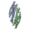
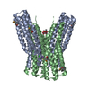
Mass: 65.409 Da / Num. of mol.: 5 / Source method: obtained synthetically / Formula: Zn
#3: Chemical
ChemComp-CL / CHLORIDE ION / Chloride


Mass: 35.453 Da / Num. of mol.: 4 / Source method: obtained synthetically / Formula: Cl
#4: Chemical ChemComp-NA / SODIUM ION


Mass: 22.990 Da / Num. of mol.: 1 / Source method: obtained synthetically / Formula: Na
#5: Chemical ChemComp-ACT / ACETATE ION / Acetate


Mass: 59.044 Da / Num. of mol.: 2 / Source method: obtained synthetically / Formula: C2H3O2
#6: Water ChemComp-HOH / water / Water


Mass: 18.015 Da / Num. of mol.: 211 / Source method: isolated from a natural source / Formula: H2O

-
Experimental details

-
Experiment

ExperimentMethod: X-RAY DIFFRACTION / Number of used crystals: 1

-
Sample preparation

CrystalDensity Matthews: 2.58 Å3/Da / Density % sol: 52.34 %
Crystal growTemperature: 298 K / Method: vapor diffusion, hanging drop / pH: 6.5
Details: Wizard II no.11 (10% 2-propanol, 0.1 M sodium cacodylate pH 6.5, 0.2 M zinc acetate), VAPOR DIFFUSION, HANGING DROP, temperature 298K

-
Data collection

DiffractionMean temperature: 100 K
Diffraction sourceSource: SYNCHROTRON / Site: APS / Beamline: 19-BM / Wavelength: 0.97903 Å
DetectorDetector: CCD / Date: Nov 23, 2010
RadiationProtocol: SINGLE WAVELENGTH / Monochromatic (M) / Laue (L): M / Scattering type: x-ray
Radiation wavelengthWavelength: 0.97903 Å / Relative weight: 1
ReflectionResolution: 2→100 Å / Num. obs: 37922 / % possible obs: 89.9 % / Observed criterion σ(I): -3 / Redundancy: 2.3 % / Rmerge(I) obs: 0.061 / Net I/σ(I): 18
Reflection shellResolution: 2→2.03 Å / Redundancy: 1.8 % / Rmerge(I) obs: 0.36 / Mean I/σ(I) obs: 2 / Num. unique all: 1313 / % possible all: 62.1

-
Processing

Software
NameVersionClassification
HKL-3000data collection
HKL-3000phasing
PHENIX(phenix.refine: 1.6.4_486)refinement
HKL-3000data reduction
HKL-3000data scaling
RefinementMethod to determine structure: SAD / Resolution: 1.997→29.882 Å / SU ML: 0.3 / Cross valid method: THROUGHOUT / σ(F): 0.94 / Phase error: 29.41 / Stereochemistry target values: ML / Details: Hydrogens were added in riding positions
RfactorNum. reflection% reflectionSelection details
Rfree0.2515 1940 5.14 %random
Rwork0.198 ---
obs0.2006 37771 88.41 %-
Solvent computationShrinkage radii: 0.89 Å / VDW probe radii: 1 Å / Solvent model: FLAT BULK SOLVENT MODEL / Bsol: 43.141 Å2 / ksol: 0.373 e/Å3
Displacement parameters
Baniso -1Baniso -2Baniso -3
1-0.7208 Å20 Å2-14.6705 Å2
2---18.0991 Å2-0 Å2
3---17.3784 Å2
Refinement stepCycle: LAST / Resolution: 1.997→29.882 Å
ProteinNucleic acidLigandSolventTotal
Num. atoms2025 0 18 211 2254
Refine LS restraints
Refine-IDTypeDev idealNumber
X-RAY DIFFRACTIONf_bond_d0.0182102
X-RAY DIFFRACTIONf_angle_d1.6572869
X-RAY DIFFRACTIONf_dihedral_angle_d14.935747
X-RAY DIFFRACTIONf_chiral_restr0.082346
X-RAY DIFFRACTIONf_plane_restr0.009384
LS refinement shell
Resolution (Å)Rfactor RfreeNum. reflection RfreeRfactor RworkNum. reflection RworkRefine-ID% reflection obs (%)
1.9969-2.06830.38051350.27272297X-RAY DIFFRACTION57
2.0683-2.15110.33181510.25533105X-RAY DIFFRACTION76
2.1511-2.24890.32381790.23683288X-RAY DIFFRACTION83
2.2489-2.36750.27221960.21893586X-RAY DIFFRACTION89
2.3675-2.51570.26681930.23133822X-RAY DIFFRACTION94
2.5157-2.70980.32672290.22243811X-RAY DIFFRACTION95
2.7098-2.98230.24212040.19863918X-RAY DIFFRACTION96
2.9823-3.41330.23512120.194021X-RAY DIFFRACTION99
3.4133-4.29840.21692200.16874007X-RAY DIFFRACTION99
4.2984-29.88550.22072210.1843976X-RAY DIFFRACTION98

+
About Yorodumi

-
News

-
Feb 9, 2022. New format data for meta-information of EMDB entries

New format data for meta-information of EMDB entries

  • Version 3 of the EMDB header file is now the official format.
  • The previous official version 1.9 will be removed from the archive.

Related info.:EMDB header

External links:wwPDB to switch to version 3 of the EMDB data model

-
Aug 12, 2020. Covid-19 info

Covid-19 info

URL: https://pdbj.org/emnavi/covid19.php

New page: Covid-19 featured information page in EM Navigator.

Related info.:Covid-19 info / Mar 5, 2020. Novel coronavirus structure data

+
Mar 5, 2020. Novel coronavirus structure data

Novel coronavirus structure data

Related info.:Yorodumi Speices / Aug 12, 2020. Covid-19 info

External links:COVID-19 featured content - PDBj / Molecule of the Month (242):Coronavirus Proteases

+
Jan 31, 2019. EMDB accession codes are about to change! (news from PDBe EMDB page)

EMDB accession codes are about to change! (news from PDBe EMDB page)

  • The allocation of 4 digits for EMDB accession codes will soon come to an end. Whilst these codes will remain in use, new EMDB accession codes will include an additional digit and will expand incrementally as the available range of codes is exhausted. The current 4-digit format prefixed with “EMD-” (i.e. EMD-XXXX) will advance to a 5-digit format (i.e. EMD-XXXXX), and so on. It is currently estimated that the 4-digit codes will be depleted around Spring 2019, at which point the 5-digit format will come into force.
  • The EM Navigator/Yorodumi systems omit the EMD- prefix.

Related info.:Q: What is EMD? / ID/Accession-code notation in Yorodumi/EM Navigator

External links:EMDB Accession Codes are Changing Soon! / Contact to PDBj

+
Jul 12, 2017. Major update of PDB

Major update of PDB

  • wwPDB released updated PDB data conforming to the new PDBx/mmCIF dictionary.
  • This is a major update changing the version number from 4 to 5, and with Remediation, in which all the entries are updated.
  • In this update, many items about electron microscopy experimental information are reorganized (e.g. em_software).
  • Now, EM Navigator and Yorodumi are based on the updated data.

External links:wwPDB Remediation / Enriched Model Files Conforming to OneDep Data Standards Now Available in the PDB FTP Archive

-
Yorodumi

Thousand views of thousand structures

  • Yorodumi is a browser for structure data from EMDB, PDB, SASBDB, etc.
  • This page is also the successor to EM Navigator detail page, and also detail information page/front-end page for Omokage search.
  • The word "yorodu" (or yorozu) is an old Japanese word meaning "ten thousand". "mi" (miru) is to see.

Related info.:EMDB / PDB / SASBDB / Comparison of 3 databanks / Yorodumi Search / Aug 31, 2016. New EM Navigator & Yorodumi / Yorodumi Papers / Jmol/JSmol / Function and homology information / Changes in new EM Navigator and Yorodumi

Read more