 Movie
Movie Controller

[English] 日本語
 Yorodumi
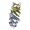
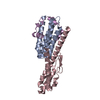
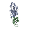
- PDB-2y7l: Structure of N-terminal domain of Candida albicans Als9-2 in comp... -
+ Open data

- Basic information
| Entry | Database: PDB / ID: 2y7l | ||||||
|---|---|---|---|---|---|---|---|
| Title | Structure of N-terminal domain of Candida albicans Als9-2 in complex with human fibrinogen gamma peptide | ||||||
 Components Components |
| ||||||
 Keywords Keywords | CELL ADHESION / ADHESIN / PEPTIDE BINDING PROTEIN | ||||||
| Function / homology |  Function and homology information Function and homology informationcell adhesion involved in multi-species biofilm formation / hyphal growth / single-species biofilm formation on inanimate substrate / yeast-form cell wall / hyphal cell wall / fibrinogen complex / Regulation of TLR by endogenous ligand / platelet alpha granule / cell adhesion involved in single-species biofilm formation / MyD88 deficiency (TLR2/4) ...cell adhesion involved in multi-species biofilm formation / hyphal growth / single-species biofilm formation on inanimate substrate / yeast-form cell wall / hyphal cell wall / fibrinogen complex / Regulation of TLR by endogenous ligand / platelet alpha granule / cell adhesion involved in single-species biofilm formation / MyD88 deficiency (TLR2/4) / positive regulation of heterotypic cell-cell adhesion / IRAK4 deficiency (TLR2/4) / plasminogen activation / MyD88:MAL(TIRAP) cascade initiated on plasma membrane / extracellular matrix structural constituent / p130Cas linkage to MAPK signaling for integrins / positive regulation of peptide hormone secretion / GRB2:SOS provides linkage to MAPK signaling for Integrins / positive regulation of exocytosis / blood coagulation, fibrin clot formation / protein polymerization / protein secretion / positive regulation of vasoconstriction / Integrin cell surface interactions / : / negative regulation of endothelial cell apoptotic process / negative regulation of extrinsic apoptotic signaling pathway via death domain receptors / fibrinolysis / side of membrane / Integrin signaling / positive regulation of substrate adhesion-dependent cell spreading / cell adhesion molecule binding / platelet alpha granule lumen / cell-matrix adhesion / positive regulation of protein secretion / Post-translational protein phosphorylation / cell-cell adhesion / Signaling by high-kinase activity BRAF mutants / MAP2K and MAPK activation / platelet activation / response to calcium ion / platelet aggregation / Regulation of Insulin-like Growth Factor (IGF) transport and uptake by Insulin-like Growth Factor Binding Proteins (IGFBPs) / Signaling by RAF1 mutants / Signaling by moderate kinase activity BRAF mutants / Paradoxical activation of RAF signaling by kinase inactive BRAF / Signaling downstream of RAS mutants / Signaling by BRAF and RAF1 fusions / Platelet degranulation / extracellular vesicle / extracellular matrix / ER-Phagosome pathway / protein-containing complex assembly / blood microparticle / positive regulation of ERK1 and ERK2 cascade / cell adhesion / endoplasmic reticulum lumen / signaling receptor binding / external side of plasma membrane / structural molecule activity / cell surface / : / extracellular exosome / extracellular region / metal ion binding / plasma membrane Similarity search - Function | ||||||
| Biological species |  CANDIDA ALBICANS (yeast) CANDIDA ALBICANS (yeast) HOMO SAPIENS (human) HOMO SAPIENS (human) | ||||||
| Method |  X-RAY DIFFRACTION / SYNCHROTRON / MOLECULAR REPLACEMENT / Resolution: 1.49 Å X-RAY DIFFRACTION / SYNCHROTRON / MOLECULAR REPLACEMENT / Resolution: 1.49 Å | ||||||
 Authors Authors | Salgado, P.S. / Cota, E. | ||||||
 Citation Citation | Journal: Proc.Natl.Acad.Sci.USA / Year: 2011 Title: Structural Basis for the Broad Specificity to Host- Cell Ligands by the Pathogenic Fungus Candida Albicans. Authors: Salgado, P.S. / Yan, R. / Taylor, J.D. / Burchell, L. / Jones, R. / Hoyer, L.L. / Matthews, S.J. / Simpson, P.J. / Cota, E. | ||||||
| History |
|
- Structure visualization
| Structure viewer | Molecule: MolmilJmol/JSmol |
|---|
- Downloads & links
-Download
| PDBx/mmCIF format | 2y7l.cif.gz | 81.1 KB | Display | PDBx/mmCIF format |
|---|---|---|---|---|
| PDB format | pdb2y7l.ent.gz | 60 KB | Display | PDB format |
| PDBx/mmJSON format | 2y7l.json.gz | Tree view | PDBx/mmJSON format | |
| Others |  Other downloads Other downloads |
-Validation report
| Arichive directory | https://data.pdbj.org/pub/pdb/validation_reports/y7/2y7lftp://data.pdbj.org/pub/pdb/validation_reports/y7/2y7l | HTTPS FTP |
|---|
-Related structure data
| Related structure data | 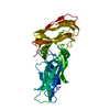 2y7mSC  2y7nC 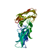 2y7oC 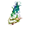 2ylhC C: citing same article ( S: Starting model for refinement |
|---|---|
| Similar structure data |


-Links
 PDBj
PDBj


- Assembly
| Deposited unit | 
| ||||||||
|---|---|---|---|---|---|---|---|---|---|
| 1 |
| ||||||||
| Unit cell |
|
-Components
| #1: Protein | Mass: 33599.988 Da / Num. of mol.: 1 / Fragment: N-TERMINAL DOMAIN, RESIDUES 18-328 Source method: isolated from a genetically manipulated source Source: (gene. exp.) CANDIDA ALBICANS (yeast) / Plasmid: PET32 XA/LIC / Production host:  |
|---|---|
| #2: Protein/peptide | Mass: 1691.780 Da / Num. of mol.: 1 / Fragment: RESIDUES 318-334 / Source method: obtained synthetically / Source: (synth.) HOMO SAPIENS (human) / References: UniProt: D3DP16, UniProt: P02679*PLUS |
| #3: Water | ChemComp-HOH /  Mass: 18.015 Da / Num. of mol.: 313 / Source method: isolated from a natural source / Formula: H2O Mass: 18.015 Da / Num. of mol.: 313 / Source method: isolated from a natural source / Formula: H2O |
| Has protein modification | Y |
| Sequence details | SEQUENCE EXCLUDES N-TERMINAL SIGNAL SEQUENCE (18 RESIDUES) FULL 17MER PEPTIDE SEQUENCE ...SEQUENCE EXCLUDES N-TERMINAL SIGNAL SEQUENCE (18 RESIDUES) FULL 17MER PEPTIDE SEQUENCE GEGQQHHLGG |
-Experimental details
-Experiment
| Experiment | Method: X-RAY DIFFRACTION / Number of used crystals: 1 |
|---|
- Sample preparation
| Crystal | Density Matthews: 2.25 Å3/Da / Density % sol: 45 % / Description: NONE |
|---|---|
| Crystal grow | pH: 8 / Details: 0.2MM AMMONIUM FLUORIDE, 20% PEG3350, PH 6.2 |
-Data collection
| Diffraction | Mean temperature: 100 K |
|---|---|
| Diffraction source | Source: SYNCHROTRON / Site: Diamond  / Beamline: I03 / Wavelength: 0.9763 / Beamline: I03 / Wavelength: 0.9763 |
| Detector | Type: ADSC CCD / Detector: CCD / Date: Nov 25, 2009 |
| Radiation | Protocol: SINGLE WAVELENGTH / Monochromatic (M) / Laue (L): M / Scattering type: x-ray |
| Radiation wavelength | Wavelength: 0.9763 Å / Relative weight: 1 |
| Reflection | Resolution: 1.49→50 Å / Num. obs: 50160 / % possible obs: 98 % / Observed criterion σ(I): 9.3 / Redundancy: 13.7 % / Biso Wilson estimate: 11.8 Å2 / Rmerge(I) obs: 0.13 / Net I/σ(I): 36.4 |
| Reflection shell | Resolution: 1.49→1.52 Å / Redundancy: 9.3 % / Rmerge(I) obs: 0.41 / Mean I/σ(I) obs: 9.3 / % possible all: 86.6 |
- Processing
| Software |
| ||||||||||||||||||||||||||||||||||||||||||||||||||||||||||||||||||||||||||||||||||||||||||||||||||||||||||||||||||||||||||||||||||||||||||||||||||||||||||||||||||||||||||||||||||||||
|---|---|---|---|---|---|---|---|---|---|---|---|---|---|---|---|---|---|---|---|---|---|---|---|---|---|---|---|---|---|---|---|---|---|---|---|---|---|---|---|---|---|---|---|---|---|---|---|---|---|---|---|---|---|---|---|---|---|---|---|---|---|---|---|---|---|---|---|---|---|---|---|---|---|---|---|---|---|---|---|---|---|---|---|---|---|---|---|---|---|---|---|---|---|---|---|---|---|---|---|---|---|---|---|---|---|---|---|---|---|---|---|---|---|---|---|---|---|---|---|---|---|---|---|---|---|---|---|---|---|---|---|---|---|---|---|---|---|---|---|---|---|---|---|---|---|---|---|---|---|---|---|---|---|---|---|---|---|---|---|---|---|---|---|---|---|---|---|---|---|---|---|---|---|---|---|---|---|---|---|---|---|---|---|
| Refinement | Method to determine structure: MOLECULAR REPLACEMENT Starting model: PDB ENTRY 2Y7M Resolution: 1.49→34.64 Å / Cor.coef. Fo:Fc: 0.945 / Cor.coef. Fo:Fc free: 0.933 / SU B: 1.222 / SU ML: 0.048 / Cross valid method: THROUGHOUT / ESU R: 0.079 / ESU R Free: 0.082 / Stereochemistry target values: MAXIMUM LIKELIHOOD / Details: HYDROGENS HAVE BEEN ADDED IN THE RIDING POSITIONS.
| ||||||||||||||||||||||||||||||||||||||||||||||||||||||||||||||||||||||||||||||||||||||||||||||||||||||||||||||||||||||||||||||||||||||||||||||||||||||||||||||||||||||||||||||||||||||
| Solvent computation | Ion probe radii: 0.8 Å / Shrinkage radii: 0.8 Å / VDW probe radii: 1.4 Å / Solvent model: MASK | ||||||||||||||||||||||||||||||||||||||||||||||||||||||||||||||||||||||||||||||||||||||||||||||||||||||||||||||||||||||||||||||||||||||||||||||||||||||||||||||||||||||||||||||||||||||
| Displacement parameters | Biso mean: 12.735 Å2
| ||||||||||||||||||||||||||||||||||||||||||||||||||||||||||||||||||||||||||||||||||||||||||||||||||||||||||||||||||||||||||||||||||||||||||||||||||||||||||||||||||||||||||||||||||||||
| Refinement step | Cycle: LAST / Resolution: 1.49→34.64 Å
| ||||||||||||||||||||||||||||||||||||||||||||||||||||||||||||||||||||||||||||||||||||||||||||||||||||||||||||||||||||||||||||||||||||||||||||||||||||||||||||||||||||||||||||||||||||||
| Refine LS restraints |
|