| Entry | Database: PDB / ID: 2y7n
|
|---|
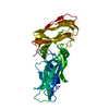
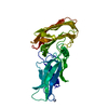
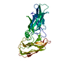
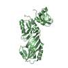
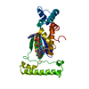
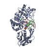
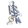
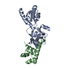
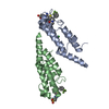
| Title | Structure of N-terminal domain of Candida albicans als9-2 - Apo Form |
|---|
 Components Components | AGGLUTININ-LIKE ALS9 PROTEIN |
|---|
 Keywords Keywords | CELL ADHESION / ADHESIN / PEPTIDE BINDING PROTEIN |
|---|
| Function / homology |  Function and homology information Function and homology information
cell adhesion involved in multi-species biofilm formation / hyphal growth / single-species biofilm formation on inanimate substrate / yeast-form cell wall / hyphal cell wall / cell adhesion involved in single-species biofilm formation / side of membrane / cell-cell adhesion / extracellular vesicle / cell adhesion ...cell adhesion involved in multi-species biofilm formation / hyphal growth / single-species biofilm formation on inanimate substrate / yeast-form cell wall / hyphal cell wall / cell adhesion involved in single-species biofilm formation / side of membrane / cell-cell adhesion / extracellular vesicle / cell adhesion / cell surface / plasma membraneSimilarity search - Function Agglutinin-like protein, N-terminal domain, N2 subdomain / Agglutinin-like protein repeat / Agglutinin-like protein, N-terminal / Agglutinin-like protein, N-terminal, N2 subdomain / Candida agglutinin-like (ALS) / Agglutinin-like protein, N-terminal domain / Cell-wall agglutinin N-terminal ligand-sugar binding / Agglutinin-like protein / Immunoglobulin-like - #1280 / Fibrogen-binding domain 1 ...Agglutinin-like protein, N-terminal domain, N2 subdomain / Agglutinin-like protein repeat / Agglutinin-like protein, N-terminal / Agglutinin-like protein, N-terminal, N2 subdomain / Candida agglutinin-like (ALS) / Agglutinin-like protein, N-terminal domain / Cell-wall agglutinin N-terminal ligand-sugar binding / Agglutinin-like protein / Immunoglobulin-like - #1280 / Fibrogen-binding domain 1 / Adhesion domain superfamily / Immunoglobulin-like / Sandwich / Mainly BetaSimilarity search - Domain/homology |
|---|
| Biological species |  CANDIDA ALBICANS (yeast) CANDIDA ALBICANS (yeast) |
|---|
| Method |  X-RAY DIFFRACTION / SYNCHROTRON / MOLECULAR REPLACEMENT / Resolution: 2 Å X-RAY DIFFRACTION / SYNCHROTRON / MOLECULAR REPLACEMENT / Resolution: 2 Å |
|---|
 Authors Authors | Salgado, P.S. / Cota, E. |
|---|
 Citation Citation | Journal: Proc.Natl.Acad.Sci.USA / Year: 2011
Title: Structural Basis for the Broad Specificity to Host- Cell Ligands by the Pathogenic Fungus Candida Albicans.
Authors: Salgado, P.S. / Yan, R. / Taylor, J.D. / Burchell, L. / Jones, R. / Hoyer, L.L. / Matthews, S.J. / Simpson, P.J. / Cota, E. |
|---|
| History | | Deposition | Jan 31, 2011 | Deposition site: PDBE / Processing site: PDBE |
|---|
| Revision 1.0 | Oct 5, 2011 | Provider: repository / Type: Initial release |
|---|
| Revision 1.1 | Oct 23, 2024 | Group: Data collection / Database references ...Data collection / Database references / Other / Structure summary
Category: chem_comp_atom / chem_comp_bond ...chem_comp_atom / chem_comp_bond / database_2 / pdbx_database_status / pdbx_entry_details / pdbx_modification_feature
Item: _database_2.pdbx_DOI / _database_2.pdbx_database_accession ..._database_2.pdbx_DOI / _database_2.pdbx_database_accession / _pdbx_database_status.status_code_sf / _pdbx_entry_details.has_protein_modification |
|---|
|
|---|
 Movie
Movie Controller

 Yorodumi
 Open data
 Basic information
 Structure visualization
 Downloads & links
 Other downloads


 PDBj
PDBj Assembly
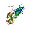

 Mass: 18.015 Da / Num. of mol.: 163 / Source method: isolated from a natural source / Formula: H2O
 Sample preparation
 / Beamline: BM14 / Wavelength: 0.9785
  Processing