 Movie
Movie Controller

[English] 日本語
 Yorodumi
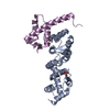
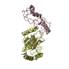
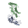
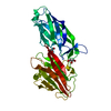
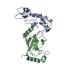
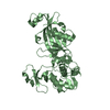
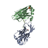
- PDB-3o6b: A Dual E3 Mechanism for Rub1 Ligation to Cdc53: Dcn1(P)-Cdc53(WHB... -
+ Open data

- Basic information
| Entry | Database: PDB / ID: 3o6b | ||||||
|---|---|---|---|---|---|---|---|
| Title | A Dual E3 Mechanism for Rub1 Ligation to Cdc53: Dcn1(P)-Cdc53(WHB) low resolution | ||||||
 Components Components |
| ||||||
 Keywords Keywords | Ligase / Cell Cycle | ||||||
| Function / homology |  Function and homology information Function and homology informationIron uptake and transport / regulation of transcription by galactose / : / cellular response to methylmercury / ubiquitin-like protein binding / positive regulation of D-glucose transmembrane transport / protein neddylation / mitochondrial fusion / ubiquitin conjugating enzyme binding / mitotic intra-S DNA damage checkpoint signaling ...Iron uptake and transport / regulation of transcription by galactose / : / cellular response to methylmercury / ubiquitin-like protein binding / positive regulation of D-glucose transmembrane transport / protein neddylation / mitochondrial fusion / ubiquitin conjugating enzyme binding / mitotic intra-S DNA damage checkpoint signaling / silent mating-type cassette heterochromatin formation / FBXL7 down-regulates AURKA during mitotic entry and in early mitosis / SCF ubiquitin ligase complex / Ubiquitin-Mediated Degradation of Phosphorylated Cdc25A / Orc1 removal from chromatin / SCF-dependent proteasomal ubiquitin-dependent protein catabolic process / Antigen processing: Ubiquitination & Proteasome degradation / DNA replication origin binding / cullin family protein binding / subtelomeric heterochromatin formation / ubiquitin ligase complex / regulation of mitotic cell cycle / G1/S transition of mitotic cell cycle / G2/M transition of mitotic cell cycle / ubiquitin-dependent protein catabolic process / protein-macromolecule adaptor activity / chromosome, telomeric region / protein ubiquitination / cell division / ubiquitin protein ligase binding / nucleus / cytoplasm Similarity search - Function | ||||||
| Biological species |  | ||||||
| Method |  X-RAY DIFFRACTION / SYNCHROTRON / MOLECULAR REPLACEMENT / Resolution: 3.1 Å X-RAY DIFFRACTION / SYNCHROTRON / MOLECULAR REPLACEMENT / Resolution: 3.1 Å | ||||||
 Authors Authors | Scott, D.C. / Monda, J.K. / Grace, C.R.R. / Duda, D.M. / Kriwacki, R.W. / Kurz, T. / Schulman, B.A. | ||||||
 Citation Citation | Journal: Mol.Cell / Year: 2010 Title: A dual E3 mechanism for Rub1 ligation to Cdc53. Authors: Scott, D.C. / Monda, J.K. / Grace, C.R. / Duda, D.M. / Kriwacki, R.W. / Kurz, T. / Schulman, B.A. | ||||||
| History |
|
- Structure visualization
| Structure viewer | Molecule: MolmilJmol/JSmol |
|---|
- Downloads & links
-Download
| PDBx/mmCIF format | 3o6b.cif.gz | 263.6 KB | Display | PDBx/mmCIF format |
|---|---|---|---|---|
| PDB format | pdb3o6b.ent.gz | 217 KB | Display | PDB format |
| PDBx/mmJSON format | 3o6b.json.gz | Tree view | PDBx/mmJSON format | |
| Others |  Other downloads Other downloads |
-Validation report
| Arichive directory | https://data.pdbj.org/pub/pdb/validation_reports/o6/3o6bftp://data.pdbj.org/pub/pdb/validation_reports/o6/3o6b | HTTPS FTP |
|---|
-Related structure data


-Links
 PDBj
PDBj


- Assembly
| Deposited unit | 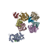
| ||||||||
|---|---|---|---|---|---|---|---|---|---|
| 1 | 
| ||||||||
| 2 | 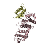
| ||||||||
| 3 | 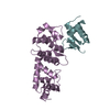
| ||||||||
| 4 | 
| ||||||||
| 5 | 
| ||||||||
| Unit cell |
|
-Components
| #1: Protein | Mass: 24353.648 Da / Num. of mol.: 5 / Fragment: DCUN1 domain, residues 70-269 Source method: isolated from a genetically manipulated source Source: (gene. exp.) Gene: DCN1, YLR128W, L3111 / Production host:  #2: Protein | Mass: 8652.943 Da / Num. of mol.: 5 / Fragment: residues 742-815 Source method: isolated from a genetically manipulated source Source: (gene. exp.) Gene: CDC53, YDL132W, D2190 / Production host: |
|---|
-Experimental details
-Experiment
| Experiment | Method: X-RAY DIFFRACTION / Number of used crystals: 1 |
|---|
- Sample preparation
| Crystal | Density Matthews: 2.59 Å3/Da / Density % sol: 52.43 % |
|---|---|
| Crystal grow | Temperature: 295 K / Method: vapor diffusion, hanging drop / pH: 7.9 Details: 21% PEG 3350, 0.1M Bis-Tris-Propane, 0.2M NaF, pH 7.9, VAPOR DIFFUSION, HANGING DROP, temperature 295K |
-Data collection
| Diffraction | Mean temperature: 77 K | |||||||||||||||||||||
|---|---|---|---|---|---|---|---|---|---|---|---|---|---|---|---|---|---|---|---|---|---|---|
| Diffraction source | Source: SYNCHROTRON / Site: APS  / Beamline: 24-ID-C / Wavelength: 0.97924 Å / Beamline: 24-ID-C / Wavelength: 0.97924 Å | |||||||||||||||||||||
| Detector | Type: ADSC QUANTUM 315 / Detector: CCD / Date: Dec 5, 2009 | |||||||||||||||||||||
| Radiation | Protocol: SINGLE WAVELENGTH / Monochromatic (M) / Laue (L): M / Scattering type: x-ray | |||||||||||||||||||||
| Radiation wavelength | Wavelength: 0.97924 Å / Relative weight: 1 | |||||||||||||||||||||
| Reflection | Resolution: 3.1→40 Å / Num. all: 30487 / Num. obs: 30204 / % possible obs: 99.1 % / Observed criterion σ(F): 0 / Observed criterion σ(I): -3 | |||||||||||||||||||||
| Reflection shell |
|
- Processing
| Software |
| ||||||||||||||||||||
|---|---|---|---|---|---|---|---|---|---|---|---|---|---|---|---|---|---|---|---|---|---|
| Refinement | Method to determine structure: MOLECULAR REPLACEMENT / Resolution: 3.1→40 Å / σ(F): 0 / Stereochemistry target values: energy minimization
| ||||||||||||||||||||
| Refinement step | Cycle: LAST / Resolution: 3.1→40 Å
|
