 Movie
Movie Controller

[English] 日本語
 Yorodumi
- PDB-5f3x: Crystal structure of Harmonin NPDZ1 in complex with ANKS4B SAM-PBM -
+ Open data

- Basic information
| Entry | Database: PDB / ID: 5f3x | ||||||
|---|---|---|---|---|---|---|---|
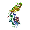
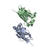
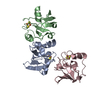
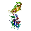
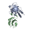
| Title | Crystal structure of Harmonin NPDZ1 in complex with ANKS4B SAM-PBM | ||||||
 Components Components |
| ||||||
 Keywords Keywords | STRUCTURAL PROTEIN/PROTEIN BINDING / COMPLEX / STRUCTURAL PROTEIN / PROTEIN BINDING / STRUCTURAL PROTEIN-PROTEIN BINDING complex | ||||||
| Function / homology |  Function and homology information Function and homology informationprotein localization to microvillus / auditory receptor cell morphogenesis / brush border assembly / stereocilia ankle link / stereocilia ankle link complex / regulation of microvillus length / parallel actin filament bundle assembly / equilibrioception / sensory perception of light stimulus / inner ear receptor cell stereocilium organization ...protein localization to microvillus / auditory receptor cell morphogenesis / brush border assembly / stereocilia ankle link / stereocilia ankle link complex / regulation of microvillus length / parallel actin filament bundle assembly / equilibrioception / sensory perception of light stimulus / inner ear receptor cell stereocilium organization / retinal cone cell development / stereocilium tip / inner ear auditory receptor cell differentiation / photoreceptor cell maintenance / stereocilium / inner ear morphogenesis / Sensory processing of sound by outer hair cells of the cochlea / Sensory processing of sound by inner hair cells of the cochlea / spectrin binding / myosin binding / microvillus / brush border / actin filament bundle assembly / photoreceptor outer segment / photoreceptor inner segment / response to endoplasmic reticulum stress / sensory perception of sound / G2/M transition of mitotic cell cycle / apical part of cell / protein-containing complex assembly / cytoskeleton / cell differentiation / cilium / synapse / endoplasmic reticulum membrane / plasma membrane / cytosol / cytoplasm Similarity search - Function | ||||||
| Biological species |  Homo sapiens (human) Homo sapiens (human) | ||||||
| Method |  X-RAY DIFFRACTION / SYNCHROTRON / MOLECULAR REPLACEMENT / Resolution: 2.649 Å X-RAY DIFFRACTION / SYNCHROTRON / MOLECULAR REPLACEMENT / Resolution: 2.649 Å | ||||||
 Authors Authors | Li, J. / He, Y. / Lu, Q. / Zhang, M. | ||||||
 Citation Citation | Journal: Dev.Cell / Year: 2016 Title: Mechanistic Basis of Organization of the Harmonin/USH1C-Mediated Brush Border Microvilli Tip-Link Complex Authors: Li, J. / He, Y. / Lu, Q. / Zhang, M. | ||||||
| History |
|
- Structure visualization
| Structure viewer | Molecule: MolmilJmol/JSmol |
|---|
- Downloads & links
-Download
| PDBx/mmCIF format | 5f3x.cif.gz | 204.1 KB | Display | PDBx/mmCIF format |
|---|---|---|---|---|
| PDB format | pdb5f3x.ent.gz | 165.1 KB | Display | PDB format |
| PDBx/mmJSON format | 5f3x.json.gz | Tree view | PDBx/mmJSON format | |
| Others |  Other downloads Other downloads |
-Validation report
| Arichive directory | https://data.pdbj.org/pub/pdb/validation_reports/f3/5f3xftp://data.pdbj.org/pub/pdb/validation_reports/f3/5f3x | HTTPS FTP |
|---|
-Related structure data
| Related structure data |  5f3yC  3k1rS C: citing same article ( S: Starting model for refinement |
|---|---|
| Similar structure data |


-Links
 PDBj
PDBj

- Assembly
| Deposited unit | 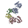
| ||||||||
|---|---|---|---|---|---|---|---|---|---|
| 1 | 
| ||||||||
| 2 | 
| ||||||||
| Unit cell |
|
-Components
| #1: Protein | Mass: 22222.609 Da / Num. of mol.: 2 / Fragment: NPDZ1, UNP residues 1-194 Source method: isolated from a genetically manipulated source Source: (gene. exp.) Homo sapiens (human) / Gene: USH1C, AIE75 / Production host:  #2: Protein | Mass: 9209.466 Da / Num. of mol.: 2 / Fragment: SAM-PBM, UNP residues 345-423 Source method: isolated from a genetically manipulated source Source: (gene. exp.) #3: Chemical |   Mass: 35.453 Da / Num. of mol.: 2 / Source method: obtained synthetically / Formula: Cl Mass: 35.453 Da / Num. of mol.: 2 / Source method: obtained synthetically / Formula: Cl#4: Water | ChemComp-HOH / |  Mass: 18.015 Da / Num. of mol.: 19 / Source method: isolated from a natural source / Formula: H2O Mass: 18.015 Da / Num. of mol.: 19 / Source method: isolated from a natural source / Formula: H2O |
|---|
-Experimental details
-Experiment
| Experiment | Method: X-RAY DIFFRACTION |
|---|
- Sample preparation
| Crystal | Density Matthews: 3.54 Å3/Da / Density % sol: 65.23 % |
|---|---|
| Crystal grow | Temperature: 289 K / Method: vapor diffusion, hanging drop / pH: 5.5 / Details: 3.0 M NaCl, 0.1 M Bis-tris |
-Data collection
| Diffraction | Mean temperature: 100 K |
|---|---|
| Diffraction source | Source: SYNCHROTRON / Site: SSRF  / Beamline: BL17U / Wavelength: 0.97915 Å / Beamline: BL17U / Wavelength: 0.97915 Å |
| Detector | Type: ADSC QUANTUM 315r / Detector: CCD / Date: Jan 14, 2015 |
| Radiation | Monochromator: Double crystal / Protocol: SINGLE WAVELENGTH / Monochromatic (M) / Laue (L): M / Scattering type: x-ray |
| Radiation wavelength | Wavelength: 0.97915 Å / Relative weight: 1 |
| Reflection | Resolution: 2.649→31.511 Å / Num. obs: 24978 / % possible obs: 99.5 % / Redundancy: 5.7 % / Net I/σ(I): 38.2 |
| Reflection shell | Resolution: 2.65→2.7 Å / Redundancy: 5.5 % / Mean I/σ(I) obs: 2.9 / % possible all: 100 |
- Processing
| Software |
| |||||||||||||||||||||||||||||||||||||||||||||||||||||||||||||||||||||||||||||||||||||||||||||||||||||||||||||||||||||||||||||||||||||||||||||||||||||||||||||||||||||||||||||||
|---|---|---|---|---|---|---|---|---|---|---|---|---|---|---|---|---|---|---|---|---|---|---|---|---|---|---|---|---|---|---|---|---|---|---|---|---|---|---|---|---|---|---|---|---|---|---|---|---|---|---|---|---|---|---|---|---|---|---|---|---|---|---|---|---|---|---|---|---|---|---|---|---|---|---|---|---|---|---|---|---|---|---|---|---|---|---|---|---|---|---|---|---|---|---|---|---|---|---|---|---|---|---|---|---|---|---|---|---|---|---|---|---|---|---|---|---|---|---|---|---|---|---|---|---|---|---|---|---|---|---|---|---|---|---|---|---|---|---|---|---|---|---|---|---|---|---|---|---|---|---|---|---|---|---|---|---|---|---|---|---|---|---|---|---|---|---|---|---|---|---|---|---|---|---|---|---|
| Refinement | Method to determine structure: MOLECULAR REPLACEMENT Starting model: 3K1R Resolution: 2.649→31.511 Å / SU ML: 0.46 / Cross valid method: FREE R-VALUE / σ(F): 1.96 / Phase error: 31.69 / Stereochemistry target values: ML
| |||||||||||||||||||||||||||||||||||||||||||||||||||||||||||||||||||||||||||||||||||||||||||||||||||||||||||||||||||||||||||||||||||||||||||||||||||||||||||||||||||||||||||||||
| Solvent computation | Shrinkage radii: 0.9 Å / VDW probe radii: 1.11 Å / Solvent model: FLAT BULK SOLVENT MODEL | |||||||||||||||||||||||||||||||||||||||||||||||||||||||||||||||||||||||||||||||||||||||||||||||||||||||||||||||||||||||||||||||||||||||||||||||||||||||||||||||||||||||||||||||
| Refinement step | Cycle: LAST / Resolution: 2.649→31.511 Å
| |||||||||||||||||||||||||||||||||||||||||||||||||||||||||||||||||||||||||||||||||||||||||||||||||||||||||||||||||||||||||||||||||||||||||||||||||||||||||||||||||||||||||||||||
| Refine LS restraints |
| |||||||||||||||||||||||||||||||||||||||||||||||||||||||||||||||||||||||||||||||||||||||||||||||||||||||||||||||||||||||||||||||||||||||||||||||||||||||||||||||||||||||||||||||
| LS refinement shell |
| |||||||||||||||||||||||||||||||||||||||||||||||||||||||||||||||||||||||||||||||||||||||||||||||||||||||||||||||||||||||||||||||||||||||||||||||||||||||||||||||||||||||||||||||
| Refinement TLS params. | Method: refined / Refine-ID: X-RAY DIFFRACTION
| |||||||||||||||||||||||||||||||||||||||||||||||||||||||||||||||||||||||||||||||||||||||||||||||||||||||||||||||||||||||||||||||||||||||||||||||||||||||||||||||||||||||||||||||
| Refinement TLS group |
|
