 Movie
Movie Controller

[English] 日本語
 Yorodumi
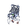
- PDB-3ivq: Structures of SPOP-Substrate Complexes: Insights into Molecular A... -
+ Open data

- Basic information
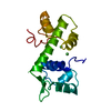
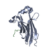
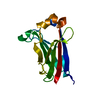
| Entry | Database: PDB / ID: 3ivq | ||||||
|---|---|---|---|---|---|---|---|
| Title | Structures of SPOP-Substrate Complexes: Insights into Molecular Architectures of BTB-Cul3 Ubiquitin Ligases: SPOPMATH-CiSBC2 | ||||||
 Components Components |
| ||||||
 Keywords Keywords | LIGASE / Protein Binding / Nucleus / Ubl conjugation pathway | ||||||
| Function / homology |  Function and homology information Function and homology informationmolecular function inhibitor activity / Cul3-RING ubiquitin ligase complex / regulation of proteolysis / Hedgehog 'on' state / protein polyubiquitination / proteasome-mediated ubiquitin-dependent protein catabolic process / nuclear speck / ubiquitin protein ligase binding / nucleoplasm / identical protein binding ...molecular function inhibitor activity / Cul3-RING ubiquitin ligase complex / regulation of proteolysis / Hedgehog 'on' state / protein polyubiquitination / proteasome-mediated ubiquitin-dependent protein catabolic process / nuclear speck / ubiquitin protein ligase binding / nucleoplasm / identical protein binding / nucleus / cytoplasm Similarity search - Function | ||||||
| Biological species |  Homo sapiens (human) Homo sapiens (human) | ||||||
| Method |  X-RAY DIFFRACTION / SYNCHROTRON / MOLECULAR REPLACEMENT / Resolution: 2.1 Å X-RAY DIFFRACTION / SYNCHROTRON / MOLECULAR REPLACEMENT / Resolution: 2.1 Å | ||||||
 Authors Authors | Schulman, B.A. / Miller, D.J. / Calabrese, M.F. / Seyedin, S. | ||||||
 Citation Citation | Journal: Mol.Cell / Year: 2009 Title: Structures of SPOP-Substrate Complexes: Insights into Molecular Architectures of BTB-Cul3 Ubiquitin Ligases. Authors: Zhuang, M. / Calabrese, M.F. / Liu, J. / Waddell, M.B. / Nourse, A. / Hammel, M. / Miller, D.J. / Walden, H. / Duda, D.M. / Seyedin, S.N. / Hoggard, T. / Harper, J.W. / White, K.P. / Schulman, B.A. | ||||||
| History |
|
- Structure visualization
| Structure viewer | Molecule: MolmilJmol/JSmol |
|---|
- Downloads & links
-Download
| PDBx/mmCIF format | 3ivq.cif.gz | 73.1 KB | Display | PDBx/mmCIF format |
|---|---|---|---|---|
| PDB format | pdb3ivq.ent.gz | 54.8 KB | Display | PDB format |
| PDBx/mmJSON format | 3ivq.json.gz | Tree view | PDBx/mmJSON format | |
| Others |  Other downloads Other downloads |
-Validation report
| Arichive directory | https://data.pdbj.org/pub/pdb/validation_reports/iv/3ivqftp://data.pdbj.org/pub/pdb/validation_reports/iv/3ivq | HTTPS FTP |
|---|
-Related structure data
| Related structure data | 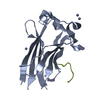 3hqhC 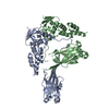 3hqiC 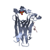 3hqlC  3hqmC  3hsvC  3htmC  3hu6C 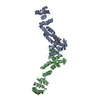 3hveC 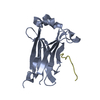 3ivvC C: citing same article ( |
|---|---|
| Similar structure data |


-Links
 PDBj
PDBj


- Assembly
| Deposited unit | 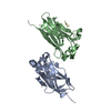
| ||||||||
|---|---|---|---|---|---|---|---|---|---|
| 1 | 
| ||||||||
| 2 | 
| ||||||||
| Unit cell |
|
-Components
| #1: Protein | Mass: 16543.967 Da / Num. of mol.: 2 Source method: isolated from a genetically manipulated source Source: (gene. exp.) Homo sapiens (human) / Gene: SPOP / Production host:  #2: Protein/peptide | Mass: 1305.371 Da / Num. of mol.: 2 / Source method: obtained synthetically #3: Water | ChemComp-HOH / |  Mass: 18.015 Da / Num. of mol.: 154 / Source method: isolated from a natural source / Formula: H2O Mass: 18.015 Da / Num. of mol.: 154 / Source method: isolated from a natural source / Formula: H2O |
|---|
-Experimental details
-Experiment
| Experiment | Method: X-RAY DIFFRACTION / Number of used crystals: 1 |
|---|
- Sample preparation
| Crystal | Density Matthews: 1.83 Å3/Da / Density % sol: 32.69 % |
|---|---|
| Crystal grow | Temperature: 298 K / Method: vapor diffusion, hanging drop / pH: 6.6 Details: 28% PME 550, 0.1 M BTP, Cryoprotection: 35% PME 550, pH 6.6, VAPOR DIFFUSION, HANGING DROP, temperature 298K |
-Data collection
| Diffraction | Mean temperature: 100 K |
|---|---|
| Diffraction source | Source: SYNCHROTRON / Site: ALS  / Beamline: 8.2.1 / Wavelength: 1 Å / Beamline: 8.2.1 / Wavelength: 1 Å |
| Radiation | Protocol: SINGLE WAVELENGTH / Monochromatic (M) / Laue (L): M / Scattering type: x-ray |
| Radiation wavelength | Wavelength: 1 Å / Relative weight: 1 |
| Reflection | Resolution: 2.1→50 Å / Num. all: 15678 / Num. obs: 15674 / % possible obs: 100 % / Observed criterion σ(F): -3 / Observed criterion σ(I): 0 / Redundancy: 7.2 % / Rsym value: 0.077 / Net I/σ(I): 26 |
| Reflection shell | Resolution: 2.1→2.18 Å / Redundancy: 7.3 % / Mean I/σ(I) obs: 4.4 / Num. unique all: 1558 / Rsym value: 0.365 / % possible all: 100 |
- Processing
| Software |
| ||||||||||||||||||||
|---|---|---|---|---|---|---|---|---|---|---|---|---|---|---|---|---|---|---|---|---|---|
| Refinement | Method to determine structure: MOLECULAR REPLACEMENT / Resolution: 2.1→40 Å / Isotropic thermal model: OVERALL ANISOTROPIC / σ(F): 0 / Stereochemistry target values: Engh & Huber
| ||||||||||||||||||||
| Displacement parameters | Biso mean: 40.6 Å2
| ||||||||||||||||||||
| Refinement step | Cycle: LAST / Resolution: 2.1→40 Å
| ||||||||||||||||||||
| Refine LS restraints |
|
