 Movie
Movie Controller

[English] 日本語
 Yorodumi
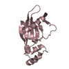
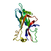
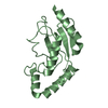
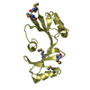
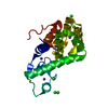
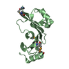
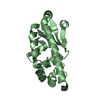
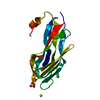
- PDB-5cw9: Crystal structure of De novo designed ferredoxin-ferredoxin domai... -
+ Open data

- Basic information
| Entry | Database: PDB / ID: 5cw9 | ||||||
|---|---|---|---|---|---|---|---|
| Title | Crystal structure of De novo designed ferredoxin-ferredoxin domain insertion protein | ||||||
 Components Components | De novo designed ferredoxin-ferredoxin domain insertion protein | ||||||
 Keywords Keywords | DE NOVO PROTEIN / designed / de novo / ferredoxin / domain insertion | ||||||
| Biological species | synthetic construct (others) | ||||||
| Method |  X-RAY DIFFRACTION / MOLECULAR REPLACEMENT / Resolution: 3.108 Å X-RAY DIFFRACTION / MOLECULAR REPLACEMENT / Resolution: 3.108 Å | ||||||
 Authors Authors | DiMaio, F. / King, I.C. / Gleixner, J. / Doyle, L. / Stoddard, B. / Baker, D. | ||||||
| Funding support |  United States, 1items United States, 1items
| ||||||
 Citation Citation | Journal: Elife / Year: 2015 Title: Precise assembly of complex beta sheet topologies from de novo designed building blocks. Authors: King, I.C. / Gleixner, J. / Doyle, L. / Kuzin, A. / Hunt, J.F. / Xiao, R. / Montelione, G.T. / Stoddard, B.L. / DiMaio, F. / Baker, D. | ||||||
| History |
|
- Structure visualization
| Structure viewer | Molecule:  MolmilJmol/JSmol MolmilJmol/JSmol |
|---|
- Downloads & links
-Download
| PDBx/mmCIF format | 5cw9.cif.gz | 41.1 KB | Display | PDBx/mmCIF format |
|---|---|---|---|---|
| PDB format | pdb5cw9.ent.gz | 28.9 KB | Display | PDB format |
| PDBx/mmJSON format | 5cw9.json.gz | Tree view | PDBx/mmJSON format | |
| Others |  Other downloads Other downloads |
-Validation report
| Arichive directory | https://data.pdbj.org/pub/pdb/validation_reports/cw/5cw9ftp://data.pdbj.org/pub/pdb/validation_reports/cw/5cw9 | HTTPS FTP |
|---|
-Related structure data


-Links
 PDBj
PDBj

- Assembly
| Deposited unit | 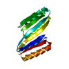
| ||||||||
|---|---|---|---|---|---|---|---|---|---|
| 1 |
| ||||||||
| Unit cell |
| ||||||||
| Details | monomer according to gel filtration |
-Components
| #1: Protein | Mass: 16838.682 Da / Num. of mol.: 1 Source method: isolated from a genetically manipulated source Source: (gene. exp.) synthetic construct (others) / Production host:  |
|---|
-Experimental details
-Experiment
| Experiment | Method: X-RAY DIFFRACTION / Number of used crystals: 1 |
|---|
- Sample preparation
| Crystal | Density Matthews: 5.36 Å3/Da / Density % sol: 77.07 % |
|---|---|
| Crystal grow | Temperature: 298 K / Method: vapor diffusion / Details: 0.1 M HEPES pH 7.5, 3.5 M NaCl |
-Data collection
| Diffraction | Mean temperature: 95 K |
|---|---|
| Diffraction source | Source: ROTATING ANODE / Type: RIGAKU MICROMAX-007 HF / Wavelength: 1.54 Å |
| Detector | Type: RIGAKU RAXIS IV++ / Detector: IMAGE PLATE / Date: Jul 31, 2014 |
| Radiation | Protocol: SINGLE WAVELENGTH / Monochromatic (M) / Laue (L): M / Scattering type: x-ray |
| Radiation wavelength | Wavelength: 1.54 Å / Relative weight: 1 |
| Reflection | Resolution: 3.1→40.282 Å / Num. obs: 6783 / % possible obs: 99.5 % / Redundancy: 13.1 % / Rmerge(I) obs: 0.077 / Net I/σ(I): 18.4 |
| Reflection shell | Resolution: 3.1→3.42 Å / Redundancy: 11.4 % / Mean I/σ(I) obs: 5.1 / % possible all: 100 |
- Processing
| Software |
| ||||||||||||||||||||||||||||||||||||||||||
|---|---|---|---|---|---|---|---|---|---|---|---|---|---|---|---|---|---|---|---|---|---|---|---|---|---|---|---|---|---|---|---|---|---|---|---|---|---|---|---|---|---|---|---|
| Refinement | Method to determine structure: MOLECULAR REPLACEMENT / Resolution: 3.108→40.282 Å / FOM work R set: 0.4538 / SU ML: 0.46 / Cross valid method: FREE R-VALUE / σ(F): 1.33 / Phase error: 53.72 / Stereochemistry target values: ML
| ||||||||||||||||||||||||||||||||||||||||||
| Solvent computation | Shrinkage radii: 0.9 Å / VDW probe radii: 1.11 Å / Solvent model: FLAT BULK SOLVENT MODEL | ||||||||||||||||||||||||||||||||||||||||||
| Displacement parameters | Biso max: 428.27 Å2 / Biso mean: 219.11 Å2 / Biso min: 154.8 Å2 | ||||||||||||||||||||||||||||||||||||||||||
| Refinement step | Cycle: final / Resolution: 3.108→40.282 Å
| ||||||||||||||||||||||||||||||||||||||||||
| Refine LS restraints |
| ||||||||||||||||||||||||||||||||||||||||||
| LS refinement shell | Refine-ID: X-RAY DIFFRACTION / Total num. of bins used: 5
|
