 Movie
Movie Controller

[English] 日本語
 Yorodumi
- PDB-1fzy: CRYSTAL STRUCTURE OF SACCHAROMYCES CEREVISIAE UBIQUITIN CONJUGATI... -
+ Open data

- Basic information
| Entry | Database: PDB / ID: 1fzy | ||||||
|---|---|---|---|---|---|---|---|
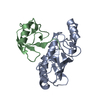
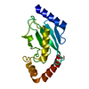
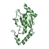
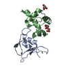
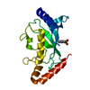
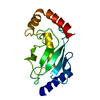
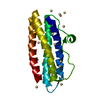
| Title | CRYSTAL STRUCTURE OF SACCHAROMYCES CEREVISIAE UBIQUITIN CONJUGATING ENZYME 1 | ||||||
 Components Components | UBIQUITIN-CONJUGATING ENZYME E2-24 KDA | ||||||
 Keywords Keywords | LIGASE / ALPHA-BETA ROLL | ||||||
| Function / homology |  Function and homology information Function and homology information: / vesicle organization / E2 ubiquitin-conjugating enzyme / proteasome binding / ubiquitin conjugating enzyme activity / Antigen processing: Ubiquitination & Proteasome degradation / ERAD pathway / protein polyubiquitination / ubiquitin-protein transferase activity / ubiquitin-dependent protein catabolic process ...: / vesicle organization / E2 ubiquitin-conjugating enzyme / proteasome binding / ubiquitin conjugating enzyme activity / Antigen processing: Ubiquitination & Proteasome degradation / ERAD pathway / protein polyubiquitination / ubiquitin-protein transferase activity / ubiquitin-dependent protein catabolic process / ATP binding / nucleus / cytosol Similarity search - Function | ||||||
| Biological species |  | ||||||
| Method |  X-RAY DIFFRACTION / Resolution: 1.9 Å X-RAY DIFFRACTION / Resolution: 1.9 Å | ||||||
 Authors Authors | Glover, M. / Williams, R.S. | ||||||
 Citation Citation | Journal: Structure / Year: 2001 Title: Structure of a conjugating enzyme-ubiquitin thiolester intermediate reveals a novel role for the ubiquitin tail. Authors: Hamilton, K.S. / Ellison, M.J. / Barber, K.R. / Williams, R.S. / Huzil, J.T. / McKenna, S. / Ptak, C. / Glover, M. / Shaw, G.S. | ||||||
| History |
|
- Structure visualization
| Structure viewer | Molecule: MolmilJmol/JSmol |
|---|
- Downloads & links
-Download
| PDBx/mmCIF format | 1fzy.cif.gz | 73.5 KB | Display | PDBx/mmCIF format |
|---|---|---|---|---|
| PDB format | pdb1fzy.ent.gz | 54.9 KB | Display | PDB format |
| PDBx/mmJSON format | 1fzy.json.gz | Tree view | PDBx/mmJSON format | |
| Others |  Other downloads Other downloads |
-Validation report
| Arichive directory | https://data.pdbj.org/pub/pdb/validation_reports/fz/1fzyftp://data.pdbj.org/pub/pdb/validation_reports/fz/1fzy | HTTPS FTP |
|---|
-Related structure data


-Links
 PDBj
PDBj


- Assembly
| Deposited unit | 
| ||||||||
|---|---|---|---|---|---|---|---|---|---|
| 1 | 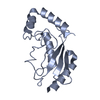
| ||||||||
| 2 | 
| ||||||||
| Unit cell |
| ||||||||
| Details | The biological assembly is a monomer. A non-crystallographically related dimer is found in the asymmetric unit. The significance of this dimer interaction is unknown. The biological assembly is a monomer. A non-crystallographically related dimer is found in the asymmetric unit. The significance of this dimer interaction is unknown. |
-Components
| #1: Protein | Mass: 16697.064 Da / Num. of mol.: 2 Source method: isolated from a genetically manipulated source Source: (gene. exp.) Production host:  #2: Water | ChemComp-HOH / |  Mass: 18.015 Da / Num. of mol.: 283 / Source method: isolated from a natural source / Formula: H2O Mass: 18.015 Da / Num. of mol.: 283 / Source method: isolated from a natural source / Formula: H2O |
|---|
-Experimental details
-Experiment
| Experiment | Method: X-RAY DIFFRACTION / Number of used crystals: 1 |
|---|
- Sample preparation
| Crystal | Density Matthews: 2.27 Å3/Da / Density % sol: 45.74 % | ||||||||||||||||||||||||||||||||||||||||||||||||||||||||||||||||||
|---|---|---|---|---|---|---|---|---|---|---|---|---|---|---|---|---|---|---|---|---|---|---|---|---|---|---|---|---|---|---|---|---|---|---|---|---|---|---|---|---|---|---|---|---|---|---|---|---|---|---|---|---|---|---|---|---|---|---|---|---|---|---|---|---|---|---|---|
| Crystal grow | Temperature: 298 K / Method: vapor diffusion, hanging drop / pH: 6 Details: PEG 5000-Monomethylether, Ammonium Sulphate, isopropanol, MES buffer, pH 6.0, VAPOR DIFFUSION, HANGING DROP, temperature 298.0K | ||||||||||||||||||||||||||||||||||||||||||||||||||||||||||||||||||
| Crystal grow | *PLUS Temperature: 20-22 ℃ / pH: 7.5 / Method: vapor diffusion / Details: used microseeding | ||||||||||||||||||||||||||||||||||||||||||||||||||||||||||||||||||
| Components of the solutions | *PLUS
|
-Data collection
| Diffraction | Mean temperature: 105 K |
|---|---|
| Diffraction source | Source: ROTATING ANODE / Type: RIGAKU RU200 / Wavelength: 1.5418 |
| Detector | Type: MAC Science DIP-2030 / Detector: IMAGE PLATE / Date: Mar 1, 1998 |
| Radiation | Protocol: SINGLE WAVELENGTH / Monochromatic (M) / Laue (L): M / Scattering type: x-ray |
| Radiation wavelength | Wavelength: 1.5418 Å / Relative weight: 1 |
| Reflection | Resolution: 1.9→20 Å / Num. all: 23300 / Num. obs: 23300 / % possible obs: 97.8 % / Observed criterion σ(F): 0 / Observed criterion σ(I): 0 / Redundancy: 3.7 % / Rmerge(I) obs: 0.101 / Net I/σ(I): 15.5 |
| Reflection shell | Resolution: 1.9→1.93 Å / Redundancy: 2.5 % / Rmerge(I) obs: 0.345 / Num. unique all: 949 / % possible all: 81.1 |
| Reflection | *PLUS Num. measured all: 85242 |
| Reflection shell | *PLUS % possible obs: 81.1 % / Mean I/σ(I) obs: 3.5 |
- Processing
| Software |
| |||||||||||||||||||||||||
|---|---|---|---|---|---|---|---|---|---|---|---|---|---|---|---|---|---|---|---|---|---|---|---|---|---|---|
| Refinement | Resolution: 1.9→20 Å / Cross valid method: FREE R-VALUE / σ(F): 0 / σ(I): 0 / Stereochemistry target values: ENGH & HUBER (CNS)
| |||||||||||||||||||||||||
| Refinement step | Cycle: LAST / Resolution: 1.9→20 Å
| |||||||||||||||||||||||||
| Refine LS restraints |
| |||||||||||||||||||||||||
| Software | *PLUS Name: CNS / Classification: refinement | |||||||||||||||||||||||||
| Refinement | *PLUS % reflection Rfree: 10 % | |||||||||||||||||||||||||
| Solvent computation | *PLUS | |||||||||||||||||||||||||
| Displacement parameters | *PLUS |
