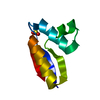
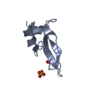
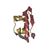
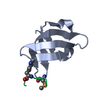
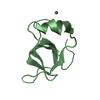
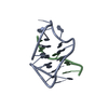
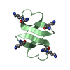
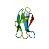
Entry Database : PDB / ID : 6q6cTitle Pore-modulating toxins exploit inherent slow inactivation to block K+ channels Kunitz-type conkunitzin-S1 Keywords Function / homology Function Domain/homology Component
/ / / / / / / / / Biological species Conus striatus (striated cone)Method / / / Resolution : 1.3 Å Authors Karbat, I. / Gueta, H. / Fine, S. / Szanto, T. / Hamer-Rogotner, S. / Dym, O. / Frolow, F. / Gordon, D. / Panyi, G. / Gurevitz, M. / Reuveny, E. Funding support Organization Grant number Country Israel Science Foundation 1248/2015
Journal : Proc.Natl.Acad.Sci.USA / Year : 2019Title : Pore-modulating toxins exploit inherent slow inactivation to block K+channels.Authors : Karbat, I. / Altman-Gueta, H. / Fine, S. / Szanto, T. / Hamer-Rogotner, S. / Dym, O. / Frolow, F. / Gordon, D. / Panyi, G. / Gurevitz, M. / Reuveny, E. History Deposition Dec 10, 2018 Deposition site / Processing site Revision 1.0 Aug 21, 2019 Provider / Type Revision 1.1 Sep 4, 2019 Group / Database references / Category / citation_authorItem _citation.country / _citation.journal_abbrev ... _citation.country / _citation.journal_abbrev / _citation.journal_id_ASTM / _citation.journal_id_CSD / _citation.journal_id_ISSN / _citation.pdbx_database_id_DOI / _citation.pdbx_database_id_PubMed / _citation.title / _citation.year / _citation_author.identifier_ORCID / _citation_author.name Revision 1.2 Sep 18, 2019 Group / Database references / Category Item / _citation.page_first / _citation.page_lastRevision 1.3 Jan 24, 2024 Group / Database references / Refinement descriptionCategory chem_comp_atom / chem_comp_bond ... chem_comp_atom / chem_comp_bond / database_2 / pdbx_initial_refinement_model / refine_hist Item / _database_2.pdbx_database_accession / _refine_hist.d_res_lowRevision 1.4 Nov 13, 2024 Group / Category / pdbx_modification_feature
Show all Show less
 Movie
Movie Controller

 Yorodumi
 Open data
 Basic information
 Components
 Keywords
 Function and homology information
 Conus striatus (striated cone)
 X-RAY DIFFRACTION /
  Authors
 Israel, 1items
  Citation
 Structure visualization
 Downloads & links
 Other downloads
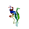


 PDBj
PDBj


 Assembly
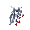
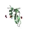


 Mass: 94.971 Da / Num. of mol.: 2 / Source method: obtained synthetically / Formula: PO4
 Mass: 62.005 Da / Num. of mol.: 6 / Source method: obtained synthetically / Formula: NO3
 Mass: 62.068 Da / Num. of mol.: 1 / Source method: obtained synthetically / Formula: C2H6O2
 Mass: 18.015 Da / Num. of mol.: 141 / Source method: isolated from a natural source / Formula: H2O
 Sample preparation
 / Beamline: ID14-4 / Wavelength: 0.98 Å
 Processing